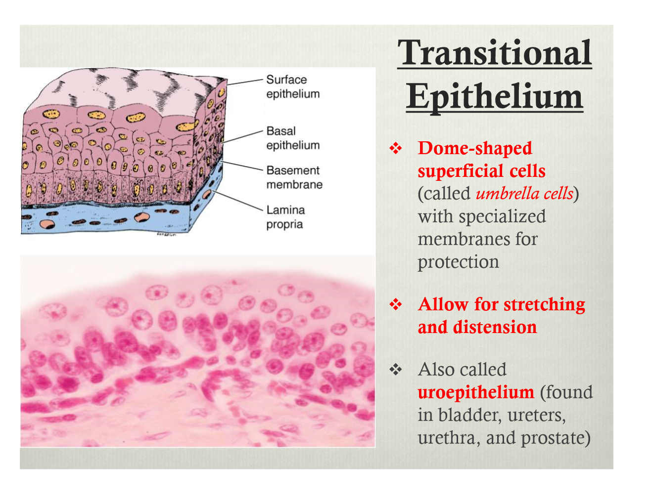
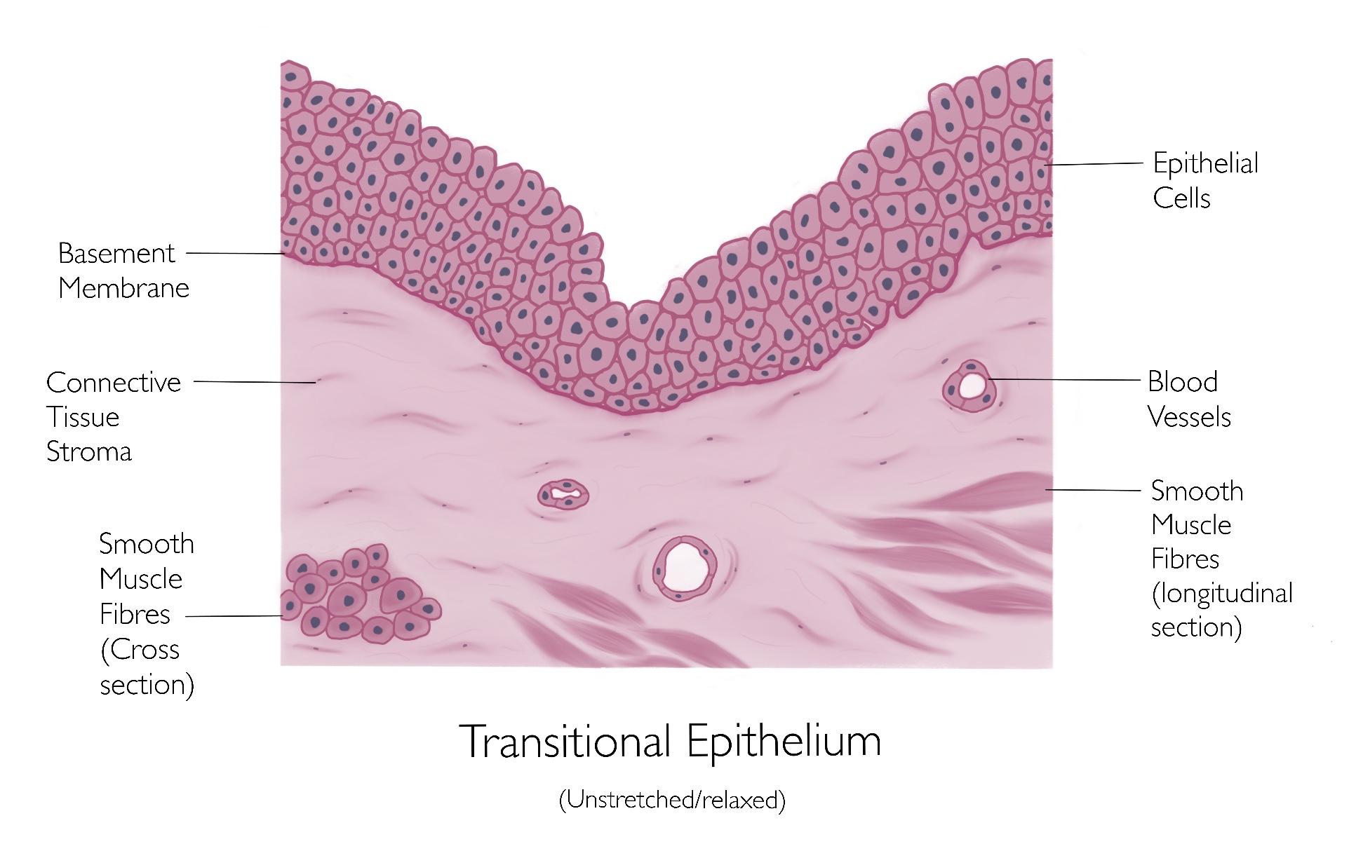
Transitional Epithelium Drawing
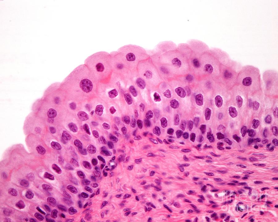
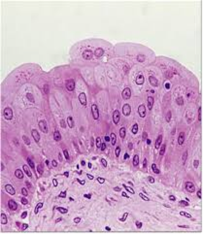
Transitional Epithelium Drawing - It rapidly adapts to distention and contraction by changing from a taller to thinner epithelium. 2.1k views 2 years ago. We will make it easy for medical students to make histology diagrams in short time during practical exams. Web you don't need an arrow for the lumen, just put the l in the lumen and that's sufficient. Web learn to draw transitional epithelium histology diagram ( for medical students) Web transitional epithelial cells are epithelial cells specialized to change shape if they are stretched laterally. Web transitional epithelium is a type of stratified epithelium composed of several layers of cells, with the morphology of cells varying depending on the function of the organ. Web transitional epithelium (urothelium) is a specialized stratified epithelium found in the lower urinary tract. Web transitional epithelium is a stratified tissue made of multiple cell layers, where the cells constituting the tissue can change shape depending on the distention in the organ. Please also make sure you lable the type of epithelium you are showing, so have a label te (transitional epithelium) and have a bracket that shows a. Web you don't need an arrow for the lumen, just put the l in the lumen and that's sufficient. Use the image slider below to learn more about the characteristics of transitional epithelium. When the tissue is stretched, the cells, especially those on the surface. Web description and photographs of transitional epithelium in the kidney and bladder, including electron micrographs. It is found only in the urinary system, specifically the ureters and urinary bladder. Web learn to draw transitional epithelium histology diagram ( for medical students) Web transitional epithelium is found only in the urinary system, specifically the ureters and urinary bladder. Transitional epithelium is a type of stratified epithelium. Learn the definition of transitional epithelial tissue and understand its. Web transitional epithelium animation, highlighting the epithelial layer, then underlying connective tissue. Web learn to draw transitional epithelium histology diagram ( for medical students) Web transitional epithelium is a type of stratified epithelium composed of several layers of cells, with the morphology of cells varying depending on the function of the organ. Web transitional epithelium is a stratified tissue made. Web epithelial tissue one layer of cube shaped cells more picky than simple squamous epi., and does not take as much wear as simple columnar epi. When the tissue is stretched, the cells, especially those on the surface. It is found only in the urinary system, specifically the ureters and urinary bladder. Empty bladder has thick folds of transitional epithelium. Web transitional epithelium is a type of stratified epithelium composed of several layers of cells, with the morphology of cells varying depending on the function of the organ. Web transitional epithelium animation, highlighting the epithelial layer, then underlying connective tissue. 2.1k views 2 years ago. Transitional epithelium is a type of stratified epithelium. Learn the definition of transitional epithelial tissue. Web transitional epithelium is a stratified tissue made of multiple cell layers, where the cells constituting the tissue can change shape depending on the distention in the organ. Web distended transitional epithelium is about two to three cell layers thick. Web you don't need an arrow for the lumen, just put the l in the lumen and that's sufficient. 2.1k. Author dan washmuth view bio. When the organ is filled with fluid, cells on the topmost layer of this epithelium can stretch and appear flattened. The image shows the wall of the urinary bladder in the relaxed state (not distended). Web transitional epithelium is a type of stratified epithelium composed of several layers of cells, with the morphology of cells. When the organ is filled with fluid, cells on the topmost layer of this epithelium can stretch and appear flattened. Good for reabsorption and secretion found kidney convoluted tubules collecting ducts ascending loop of henle choroid plexus of brain surface of ovaries thyroid gland. Make sure to include the total magnification in your key, and the objective mag. The superficial. Empty bladder has thick folds of transitional epithelium and lamina propria. Make sure to include the total magnification in your key, and the objective mag. Web epithelial tissue one layer of cube shaped cells more picky than simple squamous epi., and does not take as much wear as simple columnar epi. It is found only in the urinary system, specifically. Good for reabsorption and secretion found kidney convoluted tubules collecting ducts ascending loop of henle choroid plexus of brain surface of ovaries thyroid gland. Web distended transitional epithelium is about two to three cell layers thick. When the tissue is stretched, the cells, especially those on the surface. Web transitional epithelial cells are epithelial cells specialized to change shape if. Web transitional epithelium is a type of stratified epithelium composed of several layers of cells, with the morphology of cells varying depending on the function of the organ. 2.1k views 2 years ago. Transitional epithelium is a type of stratified epithelium. We will make it easy for medical students to make histology diagrams in short time during practical exams. Web you don't need an arrow for the lumen, just put the l in the lumen and that's sufficient. Web transitional epithelium animation, highlighting the epithelial layer, then underlying connective tissue. Web transitional epithelium (urothelium) is a specialized stratified epithelium found in the lower urinary tract. I have described how to make transitional epithelium.this is the last video of epithelium.now i will upload how to draw. It is found only in the urinary system, specifically the ureters and urinary bladder. In a relaxed state, the epithelium appears spherical or cubical, except for the apical layer, which may flatten when stretched. The superficial cells of the transitional epithelium (also called ‘umbrella cells’) are. The image shows the wall of the urinary bladder in the relaxed state (not distended). It rapidly adapts to distention and contraction by changing from a taller to thinner epithelium. It is found only in the urinary system, specifically the ureters and urinary bladder. Web transitional epithelium is an epithelial tissue which in a relaxed state appears as a stratified cuboidal epithelium. Web transitional epithelium is a stratified tissue made of multiple cell layers, where the cells constituting the tissue can change shape depending on the distention in the organ.
transitional epithelium Diagram Quizlet

Transitional Epithelium Diagram Quizlet
Urinary Bladder Transitional Epithelium 1 Photograph by Jose Calvo
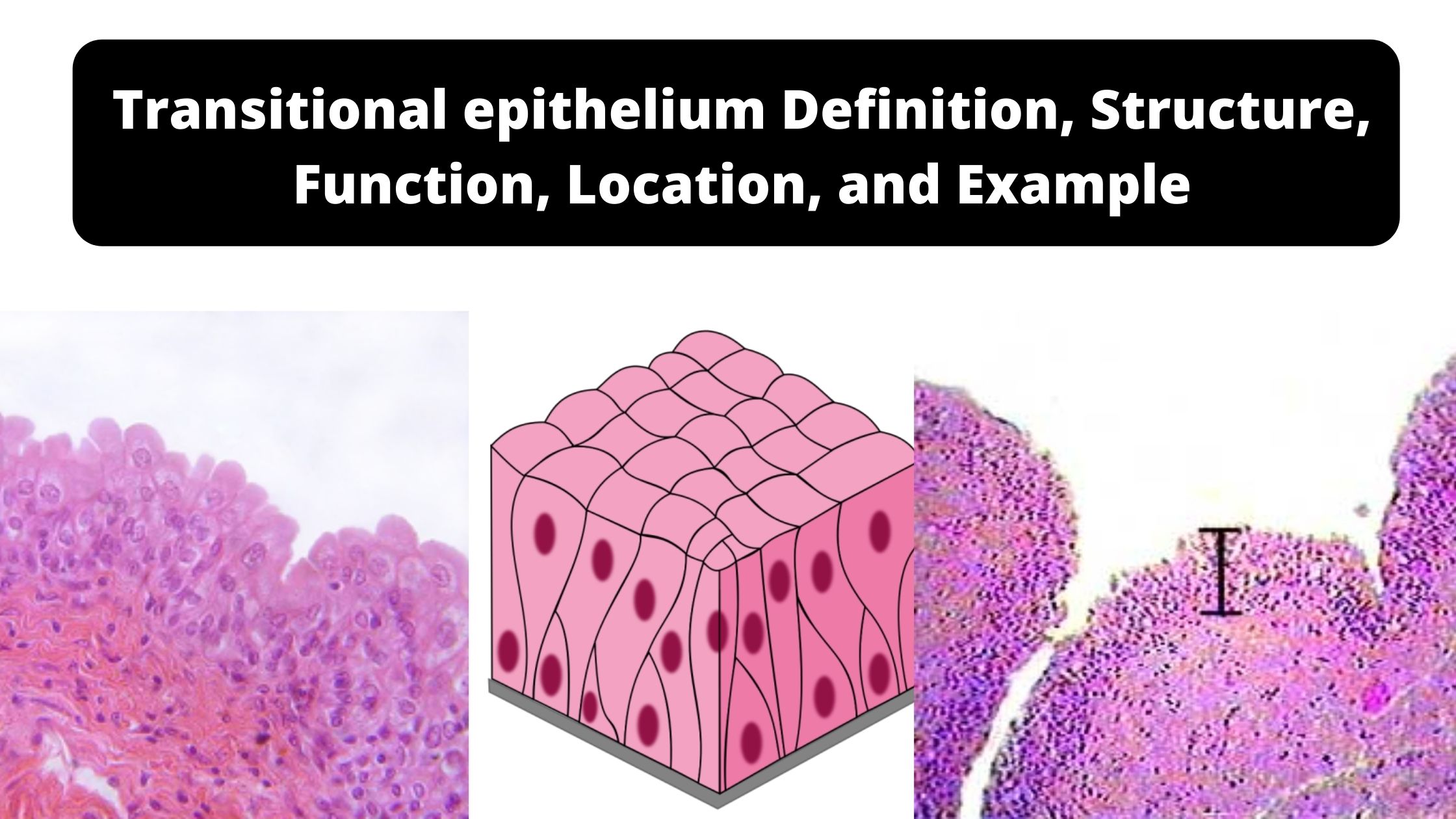
Transitional epithelium Definition, Structure, Function, Location, and
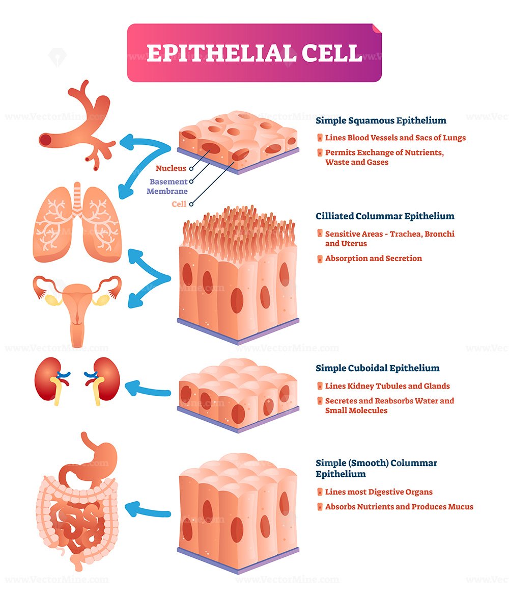
Epithelial tissue characteristics and classification scheme and types

Functions of Transitional Epithelium Tissue Video & Lesson Transcript
Transitional Epithelium
Transitional Epithelium Anatomy And Physiology Histol vrogue.co
Epithelial Tissues Anatomy 101
Epithelial cells vector illustration VectorMine
323 Views 2 Years Ago.
Author Dan Washmuth View Bio.
It Is Found Only In The Urinary System, Specifically The Ureters And Urinary Bladder.
Web Learn To Draw Transitional Epithelium Histology Diagram ( For Medical Students)
Related Post: