Muscle Cell Drawing
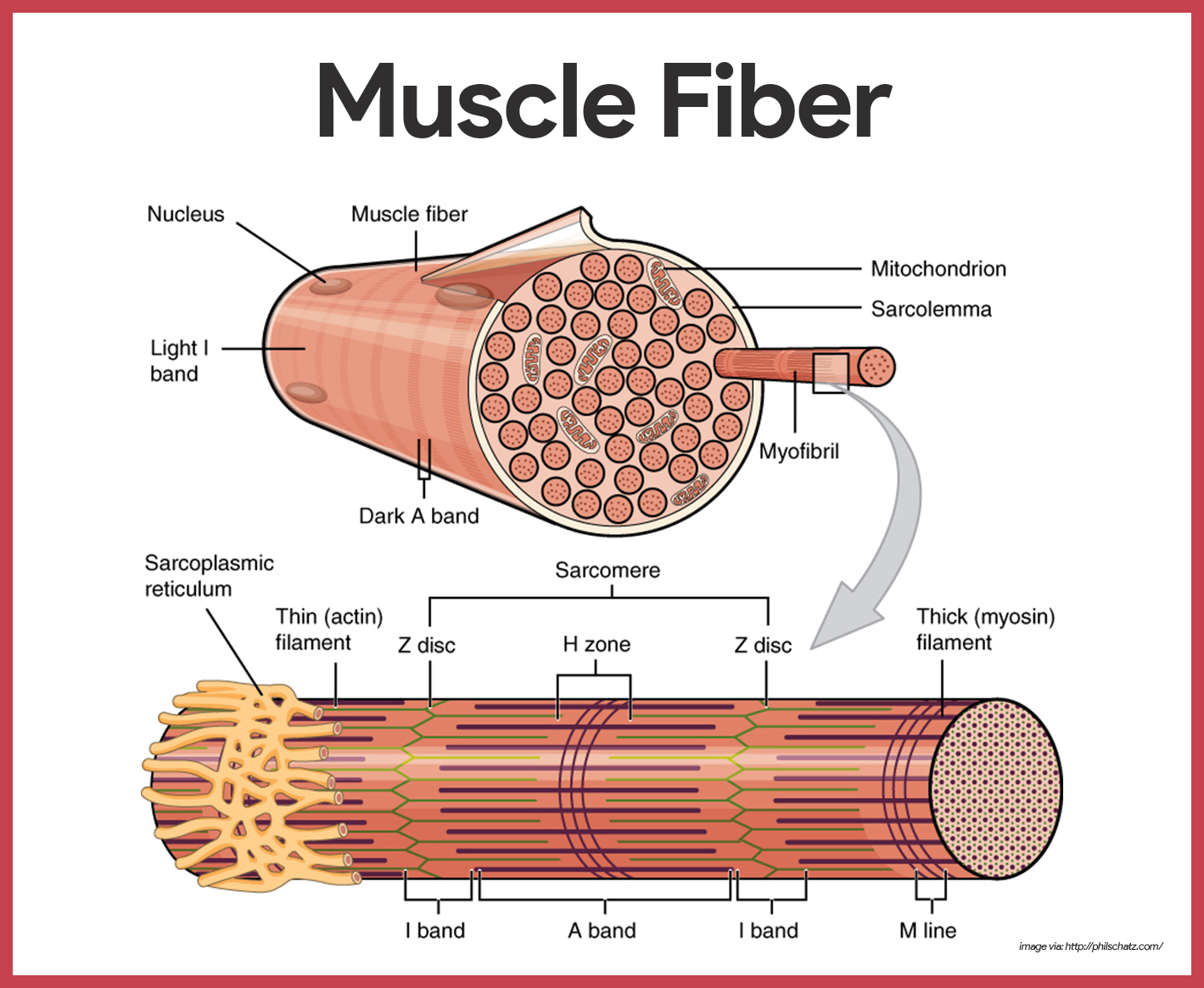
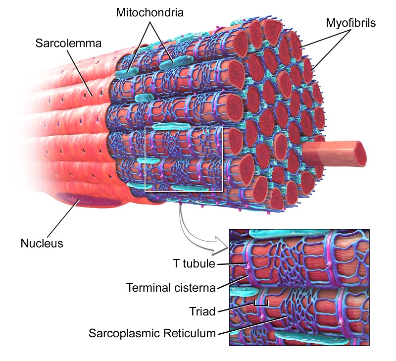
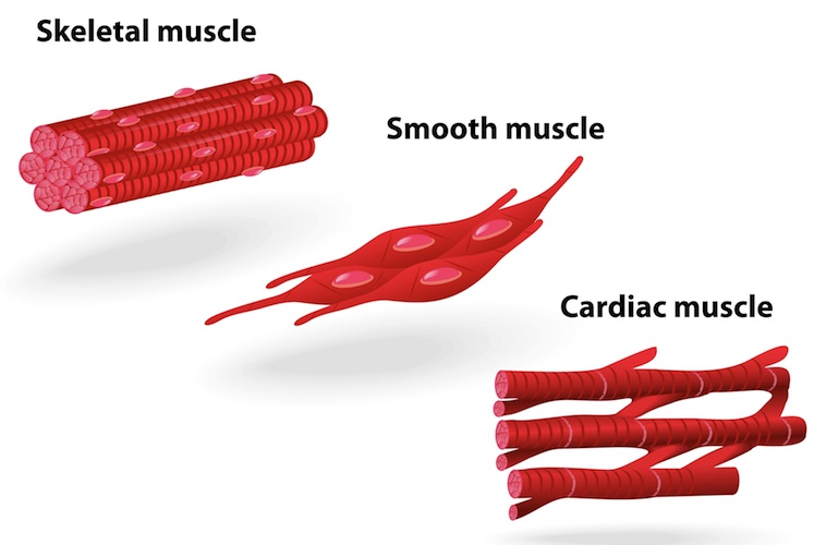
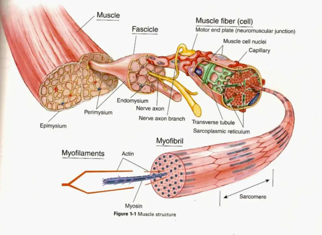
Muscle Cell Drawing - View muscle cell drawing videos. While several associated proteins help, actin and myosin form thick and thin filaments which slide past each other to contract small units of a muscle cell. Cardiac muscle is involuntary and found only in the heart. Web this is what we wanted to get to, but we're going to go even within the muscle cell to see, understand how all the myosin and the actin filaments fit into that muscle cell. Central to this contraction mechanism are the proteins actin and myosin, which form the thick and thin filaments, respectively. Skeletal muscle is voluntary and responds to conscious stimuli. Web how to draw a cell. Web human body maps. The cytoplasm is homogeneously eosinophilic and consists mainly of myofilaments. Cardiac muscle cell and centered nucleus. With this, you can now add color to your cell drawing; Learn about the three types of muscle as you use our 3d models to explore the anatomical structure and physiology of human muscles. Web human body maps. Web muscle cells, scientifically termed myocytes, are specialized cells in animals designed for contraction. Muscular system, which includes all types of muscles. Skeletal muscle is voluntary and responds to conscious stimuli. Web table of contents. Web the structure of a muscle cell can be explained using a diagram labelling muscle filaments, myofibrils, sarcoplasm, cell nuclei (nuclei is the plural word for the singular nucleus), sarcolemma, and the fascicle of which the muscle fibre is part. Muscle cell drawing pictures, images and stock. Web muscle cells, scientifically termed myocytes, are specialized cells in animals designed for contraction. Web because skeletal muscle cells are long and cylindrical, they are commonly referred to as muscle fibers. Learn this topic now at kenhub! Describe the structure and function of skeletal muscle fibers. The cells are striated and multinucleated appearing as long, unbranched cylinders. Skeletal muscle fibers can be quite large for human cells, with diameters up to 100 μ m and lengths up to 30 cm (11.8 in) in the sartorius of the upper leg. Web human body maps. Muscular system, which includes all types of muscles in the body. The process of muscle contraction begins at the site where a motor neuron’s. Web this article describes the histology of the muscle cells types: This article is about skeletal myocytes. The various shapes are called ribosomes, lysogens and vacuoles. Muscle cell drawing pictures, images and stock photos. Muscle cells are commonly called myocytes. Web muscle cell diagram. Scribble around inside the cell with wavy lines are ovals. Learn about the three types of muscle as you use our 3d models to explore the anatomical structure and physiology of human muscles. Web because skeletal muscle cells are long and cylindrical, they are commonly referred to as muscle fibers. Muscle cell drawing pictures, images and. It is subdivided into two broad systems: The various shapes are called ribosomes, lysogens and vacuoles. Contractile tissue is able to generate tension of force. Web human body maps. View muscle cell drawing videos. The cytoplasm is homogeneously eosinophilic and consists mainly of myofilaments. Every skeletal muscle fiber in every skeletal muscle is innervated by a motor neuron at a nmj. Skeletal muscle and cardiac muscle. Cardiac muscle cell and centered nucleus. While several associated proteins help, actin and myosin form thick and thin filaments which slide past each other to contract small units. Web how to draw a cell. Excitable tissue responds to stimuli through electrical signals. Skeletal muscle fibers can be quite large for human cells, with diameters up to 100 μ m and lengths up to 30 cm (11.8 in) in the sartorius of the upper leg. Web the structure of a muscle cell can be explained using a diagram labelling. Muscle cell drawing pictures, images and stock photos. Contractile tissue is able to generate tension of force. Web the musculoskeletal system (locomotor system) is a human body system that provides our body with movement, stability, shape, and support. A muscle cell, also known as a myocyte, is a mature contractile cell in the muscle of an animal. Myocytes, sometimes called. Muscle cell drawing pictures, images and stock photos. In humans and other vertebrates there are three types: Cytology of skeletal muscle cells. Web the musculoskeletal system (locomotor system) is a human body system that provides our body with movement, stability, shape, and support. Cardiac muscle microscope slide identification. Cardiac muscle is involuntary and found only in the heart. Skeletal, smooth and cardiac muscle cells. So this right here is a muscle cell or a myofiber. Web how to draw a cell. The cardiac muscle under the microscope. These cells possess a unique arrangement of motor proteins that enable them to reduce their length. Within muscles, there are layers of connective tissue called the epimysium, perimysium, and endomysium. Unique features of cardiac muscle in a light microscope. Web because skeletal muscle cells are long and cylindrical, they are commonly referred to as muscle fibers. A muscle cell, also known as a myocyte, is a mature contractile cell in the muscle of an animal. Every skeletal muscle fiber in every skeletal muscle is innervated by a motor neuron at a nmj.
Diagram showing types of muscle cells illustration Stock Vector Image
Muscular System Anatomy and Physiology Nurseslabs

Types of muscle cells vector illustration in 2022 Types of muscles
Muscle Cell (Myocyte) Definition, Function & Structure Biology
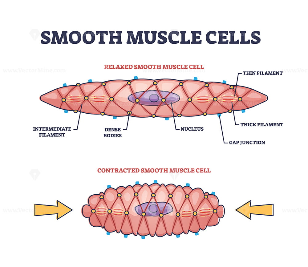
Smooth Muscle Cell Structure

How To Draw Muscle Cell Step by Step YouTube
muscle tissue types diagram

Skeletal Muscle Cell Structure
Muscle cell diagram

Types of muscle cell diagram 1783902 Vector Art at Vecteezy
Excitable Tissue Responds To Stimuli Through Electrical Signals.
Learn About The Three Types Of Muscle As You Use Our 3D Models To Explore The Anatomical Structure And Physiology Of Human Muscles.
Scribble Around Inside The Cell With Wavy Lines Are Ovals.
The Cytoplasm Is Homogeneously Eosinophilic And Consists Mainly Of Myofilaments.
Related Post: