Labeled Heart Drawing
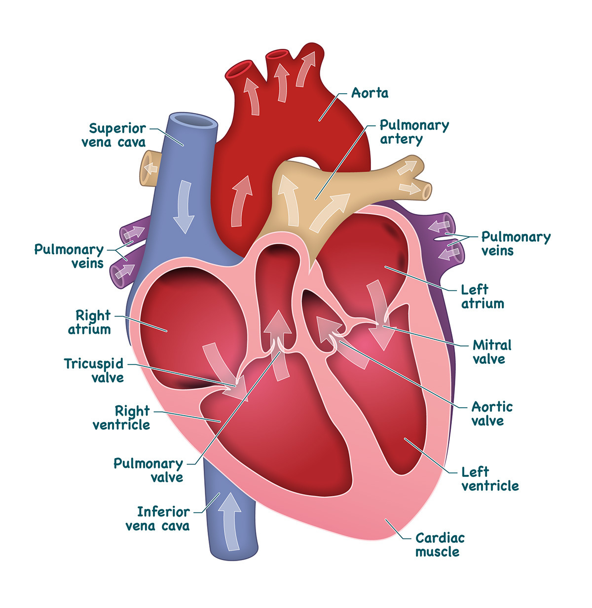
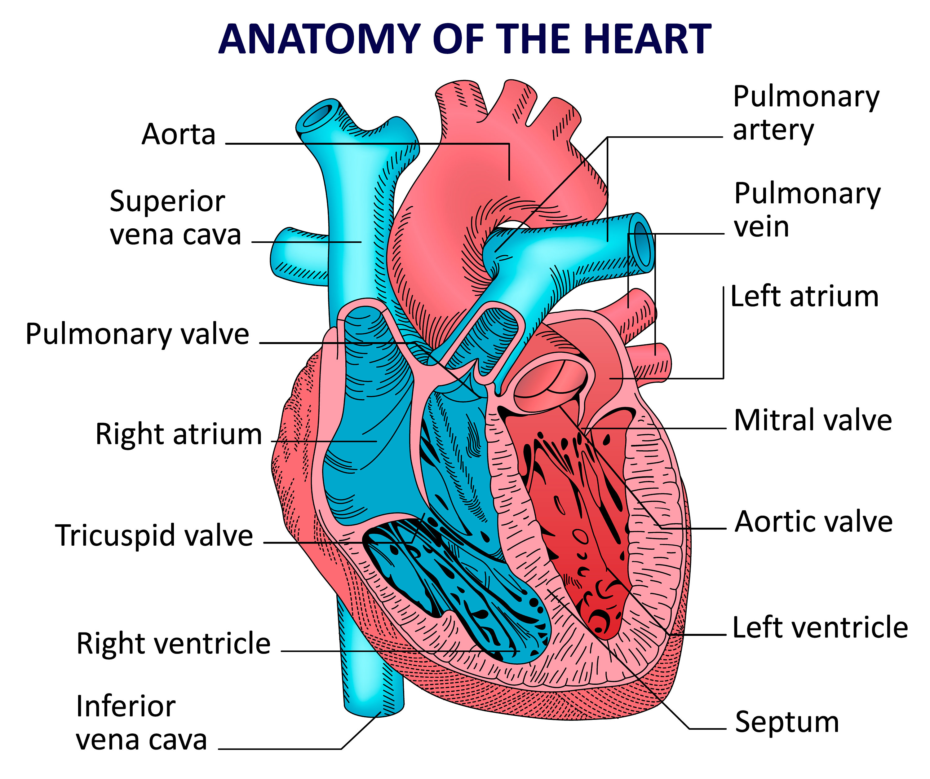
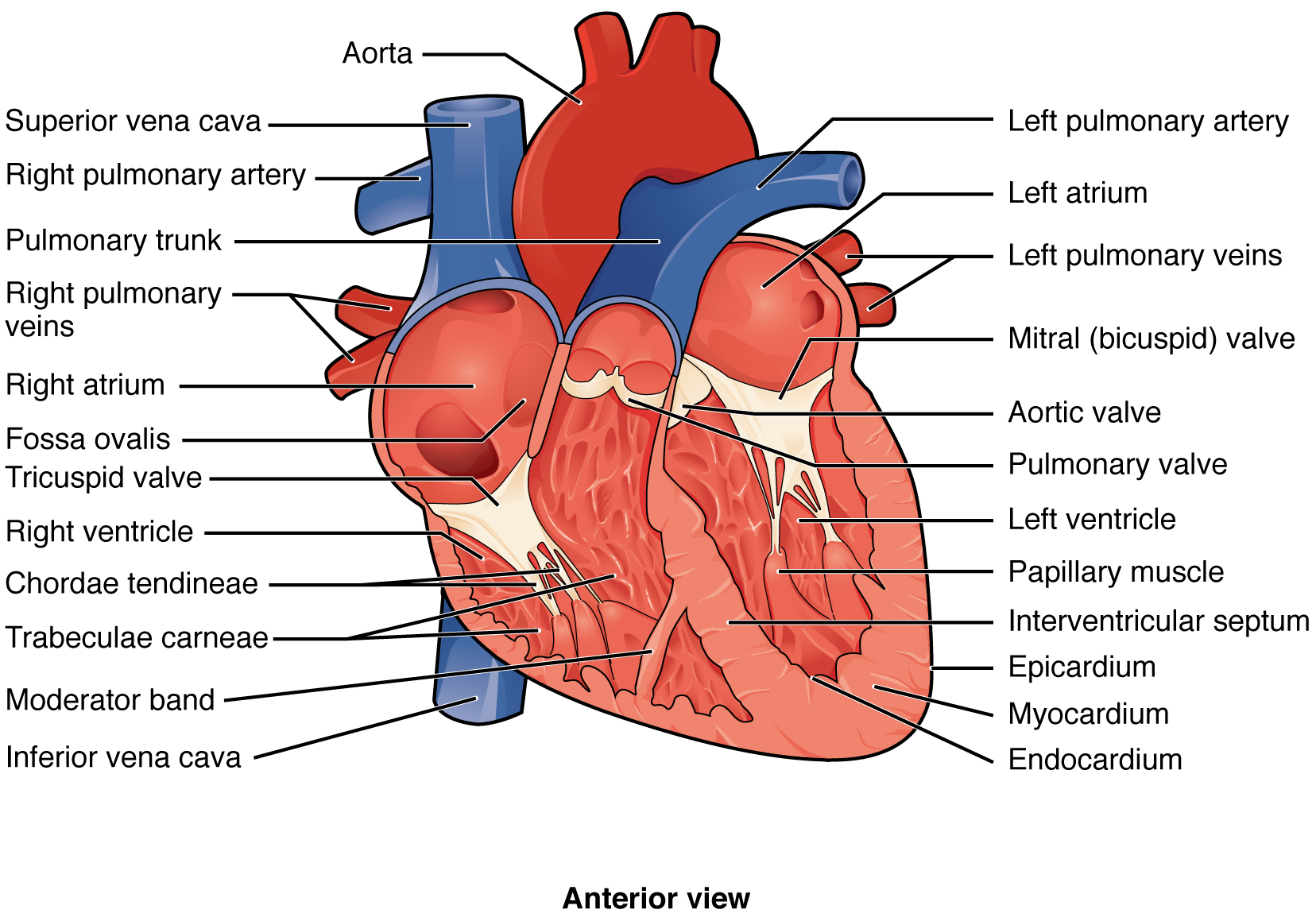
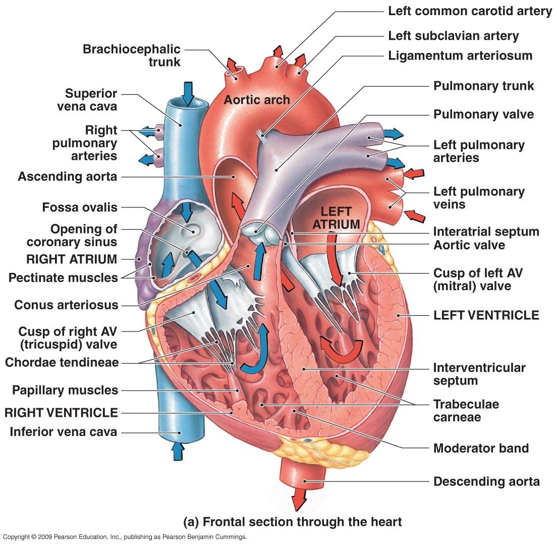
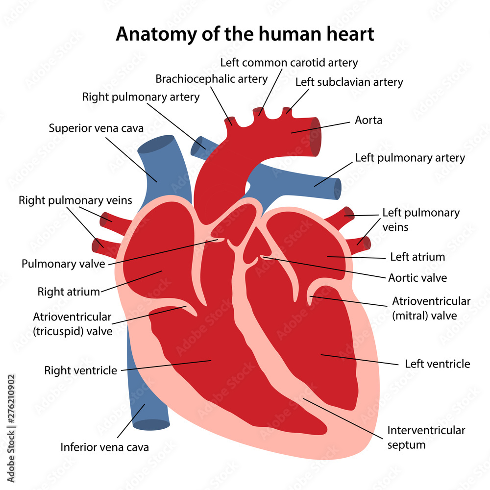
Labeled Heart Drawing - The heart features four types of valves which regulate the flow of blood through the heart. It consists of four chambers, four valves, two main arteries (the coronary arteries), and the conduction system. The video above also provides an animation at the end to quiz yourself and test your knowledge! Learn more about the heart in this article. Difference between arteries and veins. Rotate the 3d model to see how the heart's valves control blood flow between heart chambers and blood flow out of the heart. Learn all about the heart, blood vessels, and composition of blood itself with our 3d models and explanations of cardiovascular system anatomy and physiology. Web your heart’s main function is to move blood throughout your body. The heart is a muscular organ situated in the mediastinum. Web inside, the heart is divided into four heart chambers: Web label the parts of the heart to reference it for anatomy. The left and right sides of the heart have different functions: You can also refer to the byju’s app for further reference. Web 1.3m views 3 years ago 3 products. [right atrium and ventricle of the heart (labeled)] Explore the diagram of heart along with the structural details only at byju’s. You can also refer to the byju’s app for further reference. The video above also provides an animation at the end to quiz yourself and test your knowledge! Describe the location and position of the heart within the body cavity. Anatomy of the heart pericardium. See the labelling the heart activity for additional support in using this interactive. Blood brings oxygen and nutrients to your cells. Web labeled heart diagrams. Labeled heart diagram showing the heart from anterior. Learn all about the heart, blood vessels, and composition of blood itself with our 3d models and explanations of cardiovascular system anatomy and physiology. Anatomy of the heart pericardium. Web inside, the heart is divided into four heart chambers: Web selecting or hovering over a box will highlight each area in the diagram. [right atrium and ventricle of the heart (labeled)] It also takes away carbon dioxide and other waste so other organs can dispose of them. Web how to draw the internal structure of the heart. They permit blood flow in one direction only, and prevent backflow of blood. Anatomy of the heart made easy along with the blood flow through the cardiac structures, valves, atria, and ventricles. The four types of valves are: Web 1.3m views 3 years ago 3 products. Web the cardiovascular system. If you're trying to identify parts of the heart for a class or just for fun, consider adding the names of each segment. The heart is a mostly hollow, muscular organ composed of cardiac muscles and connective tissue that acts as a pump to distribute blood throughout the body’s tissues. Once you’re feeling confident, you can. They permit blood flow in one direction only, and prevent backflow of blood. Blood brings oxygen and nutrients to your cells. Rotate the 3d model to see how the heart's valves control blood flow between heart chambers and blood flow out of the heart. Web function and anatomy of the heart made easy using labeled diagrams of cardiac structures and. See the labelling the heart activity for additional support in using this interactive. New 3d rotate and zoom. Web anatomy of the human heart and coronaries: Web anatomy of the human heart. Blood flow through the heart, cardiac circulation pathway, and anatomy of the heart. New 3d rotate and zoom. It also takes away carbon dioxide and other waste so other organs can dispose of them. It consists of four chambers, four valves, two main arteries (the coronary arteries), and the conduction system. By the end of this section, you will be able to: [right atrium and ventricle of the heart (labeled)] Anatomy of the heart made easy along with the blood flow through the cardiac structures, valves, atria, and ventricles. How to visualize anatomic structures. The heart features four types of valves which regulate the flow of blood through the heart. Identify the tissue layers of the heart; Web in just a few minutes, you will be able to label the. These valves have been clearly shown in the labeled diagram of the heart. Once you’re feeling confident, you can test yourself using the unlabeled diagrams of the parts of the heart below. Relate the structure of the heart to its function as a pump; Controls the rhythm and speed of your heart rate. By the end of this section, you will be able to: Blood flow through the heart, cardiac circulation pathway, and anatomy of the heart. Rotate the 3d model to see how the heart's valves control blood flow between heart chambers and blood flow out of the heart. Compare systemic circulation to pulmonary circulation; Anatomy of the heart pericardium. If you're trying to identify parts of the heart for a class or just for fun, consider adding the names of each segment. Web label the parts of the heart to reference it for anatomy. Web in just a few minutes, you will be able to label the entire diagram shown below! For optimal viewing of this interactive, view at your screen’s default zoom setting (100%) and with your browser window view maximised. New 3d rotate and zoom. Neatly print the names around your drawing and then use a ruler to draw an arrow to the corresponding part. Web your heart’s main function is to move blood throughout your body.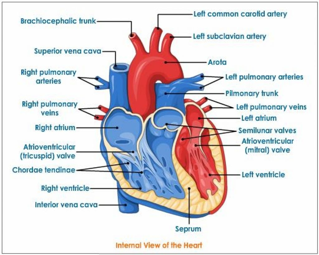
Heart And Labels Drawing at GetDrawings Free download
Heart And Labels Drawing at GetDrawings Free download
Human heart anatomy. Vector diagram Etsy
Heart Anatomy Labeled Diagram, Structures, Blood Flow, Function of

heart diagram labeled Vector illustration of diagram of human heart

31 Human Heart To Label Labels Design Ideas 2020

How to Draw the Internal Structure of the Heart (with Pictures)
OpenStax AnatPhys fig.19.9 Internal Anatomy of the Heart English
Labeled Drawing Of The Heart at GetDrawings Free download
Anatomy of the human heart. Cross sectional diagram of the heart with
The Video Above Also Provides An Animation At The End To Quiz Yourself And Test Your Knowledge!
Difference Between Arteries And Veins.
Blood Brings Oxygen And Nutrients To Your Cells.
See The Labelling The Heart Activity For Additional Support In Using This Interactive.
Related Post:
