Human Heart Drawing Labeled
Human Heart Drawing Labeled - The upper two chambers of the heart are called auricles. How to visualize anatomic structures. Begin by sketching a rounded, lumpy, irregular figure. Figure 19.2 shows the position of the heart within the thoracic cavity. Do you want a fun way to learn the structure of the heart? Next you will draw the aortic arch. This outlines the lower chamber of the heart, which includes both the left and right ventricles. Web practise labelling the human heart diagram. It consists of four chambers, four valves, two main arteries (the coronary arteries), and the conduction system. The chambers are separated by heart valves, which make sure that the blood keeps flowing in the right direction. Within the mediastinum, the heart is separated from the other mediastinal structures by a tough membrane known as the pericardium, or pericardial sac. June 20, 2023 fact checked. It’s your circulatory system ’s main organ. Web drawing a human heart is easier than you may think. Learn more about the heart in this article. Next you will draw the aortic arch. This key circulatory system structure is comprised of four chambers. Web inside, the heart is divided into four heart chambers: It has four hollow chambers surrounded by muscle and other heart tissue. How to visualize anatomic structures. Images are labelled, providing an invaluable medical and anatomical tool. Web drawing a human heart is easier than you may think. It should look a bit like the shape of africa. Introduction to the human heart. The heart is a muscular organ situated in the mediastinum. Images are labelled, providing an invaluable medical and anatomical tool. Two atria (right and left) and two ventricles (right and left). This tool provides access to several medical illustrations, allowing the user to interactively discover heart anatomy. The upper two chambers of the heart are called auricles. Your heart is a muscular organ that pumps blood to your body. Are you fascinated with anatomy, or looking to take your drawing skills to the next level? Extend two curved lines upwards from the irregular shape. It’s your circulatory system ’s main organ. Plus, you may just learn something new along the way. Web practise labelling the human heart diagram. After all, we know that stress is bad for the heart! June 20, 2023 fact checked. The heart is a muscular organ situated in the mediastinum. Web heart pictures, diagram & anatomy | body maps. The heart is a hollow, muscular organ that pumps oxygenated blood throughout the body and deoxygenated blood to the lungs. It is about the size of a fist, is broad at the top, and tapers toward the base. Read more about heart valves and how they help blood flow through the heart. The heart—the primary organ of the cardiovascular system—is a muscle that contracts regularly, via a natural pacemaker that produces electrical impulses. The chambers are separated by heart valves,. The heart is a mostly hollow, muscular organ composed of cardiac muscles and connective tissue that acts as a. The heart is located within the thoracic cavity, medially between the lungs in the mediastinum. 244 × 240 pixels | 489 × 480 pixels | 782 × 768 pixels | 1,043 × 1,024 pixels | 2,086 × 2,048 pixels | 663. Web function and anatomy of the heart made easy using labeled diagrams of cardiac structures and blood flow through the atria, ventricles, valves, aorta, pulmonary arteries veins, superior inferior vena cava, and chambers. It’s your circulatory system ’s main organ. June 20, 2023 fact checked. The upper two chambers of the heart are called auricles. Do you want a fun. Web drawing a human heart is easier than you may think. Drag and drop the text labels onto the boxes next to the diagram. Size of this png preview of this svg file: Web heart, organ that serves as a pump to circulate the blood. Read more about heart valves and how they help blood flow through the heart. Do you want a fun way to learn the structure of the heart? Web 1 finding a diagram. The upper two chambers of the heart are called auricles. Web the human heart is located within the thoracic cavity, medially between the lungs in the space known as the mediastinum. Next you will draw the aortic arch. Begin by sketching a rounded, lumpy, irregular figure. Selecting or hovering over a box will highlight each area in the diagram. The heart is a muscular organ situated in the mediastinum. It is about the size of a fist, is broad at the top, and tapers toward the base. June 20, 2023 fact checked. Extend two curved lines upwards from the irregular shape. Learn more about the heart in this article. Size of this png preview of this svg file: This outlines the lower chamber of the heart, which includes both the left and right ventricles. The size of the heart is the size of about a clenched fist. Drag and drop the text labels onto the boxes next to the diagram.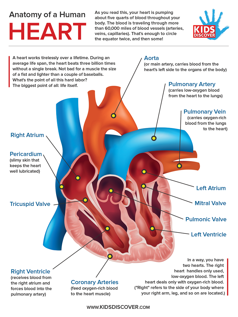
Infographic Anatomy of the Human Heart Kids Discover

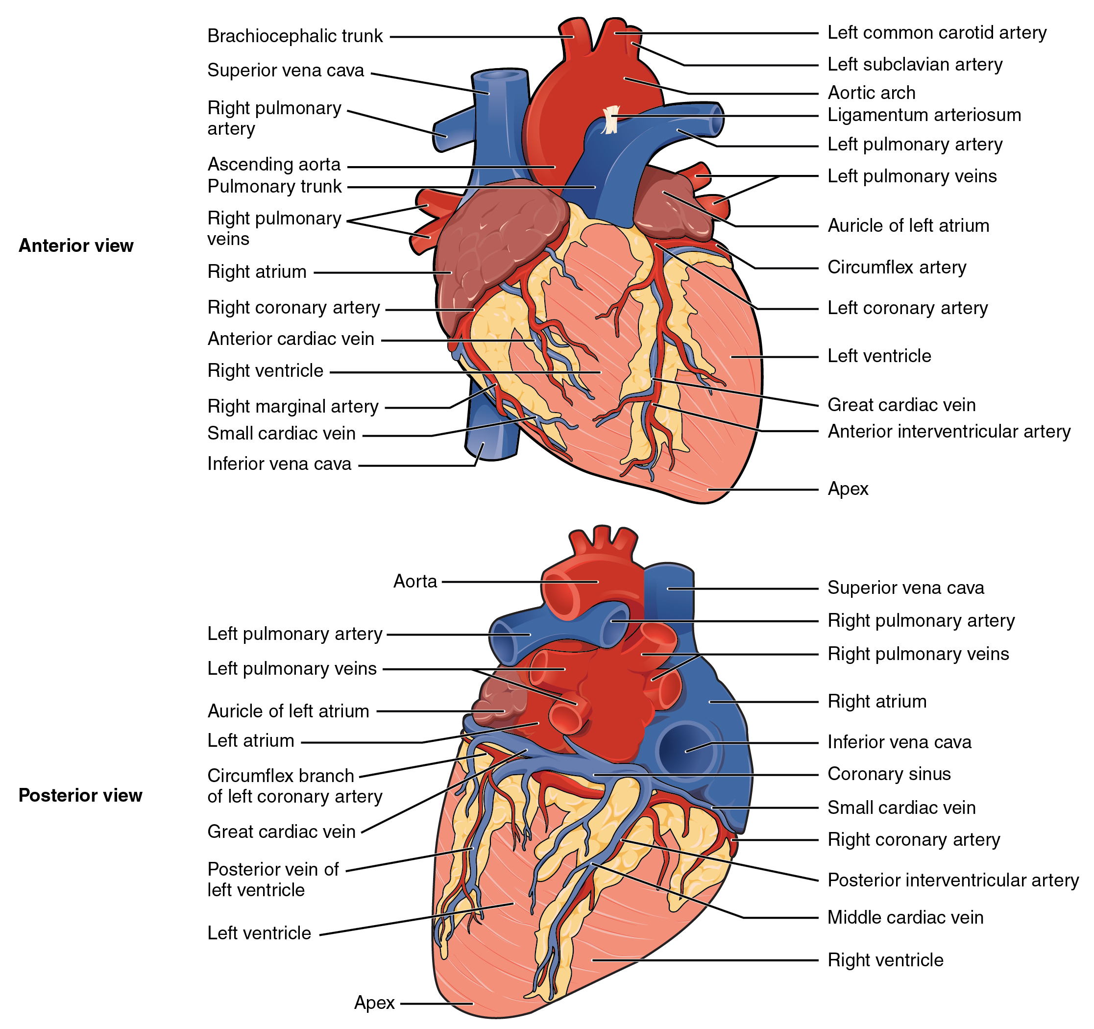
External Structure Of Heart Anatomy Diagram
19.1 Heart Anatomy Anatomy and Physiology
Heart Anatomy Labeled Diagram, Structures, Blood Flow, Function of

Heart Anatomy chambers, valves and vessels Anatomy & Physiology

humanheartdiagram Tim's Printables

How to Draw the Internal Structure of the Heart 14 Steps
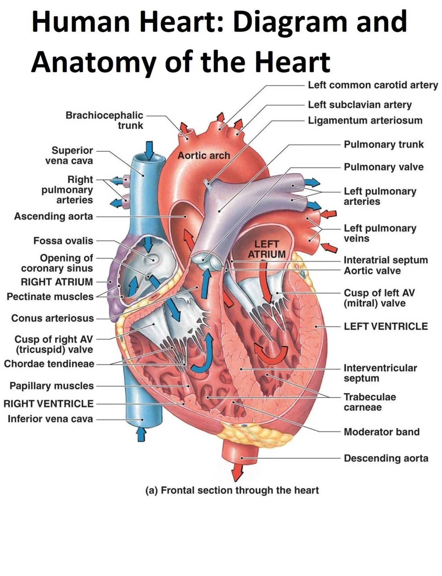
The Human Heart Diagram Display Poster Diagram and Anatomy of the Heart
.svg/1043px-Diagram_of_the_human_heart_(cropped).svg.png)
FileDiagram of the human heart (cropped).svg Wikipedia

Human heart anatomy. Vector diagram in 2021 Heart anatomy, Human
Your Heart Is In The Center Of Your Chest, Near Your Lungs.
Web Heart Pictures, Diagram & Anatomy | Body Maps.
The Heart Is A Hollow, Muscular Organ That Pumps Oxygenated Blood Throughout The Body And Deoxygenated Blood To The Lungs.
Web Practise Labelling The Human Heart Diagram.
Related Post:
