Heart Anatomy Draw
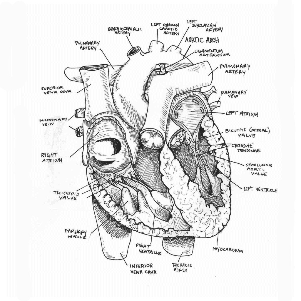
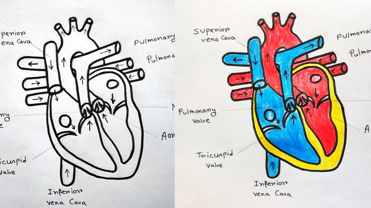
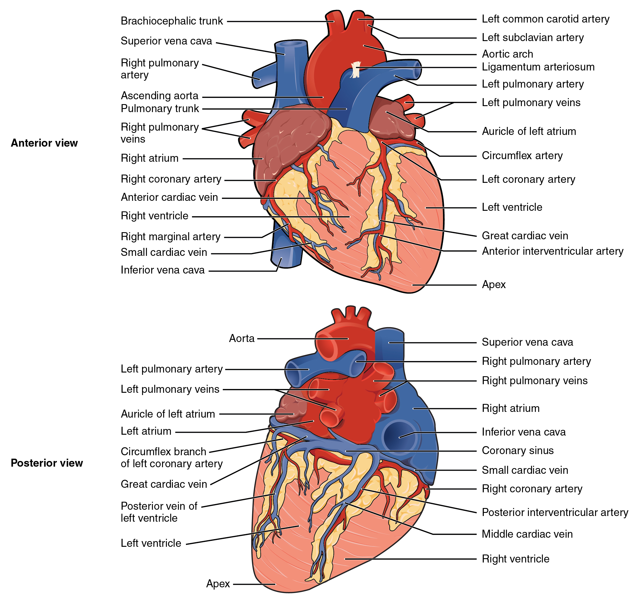
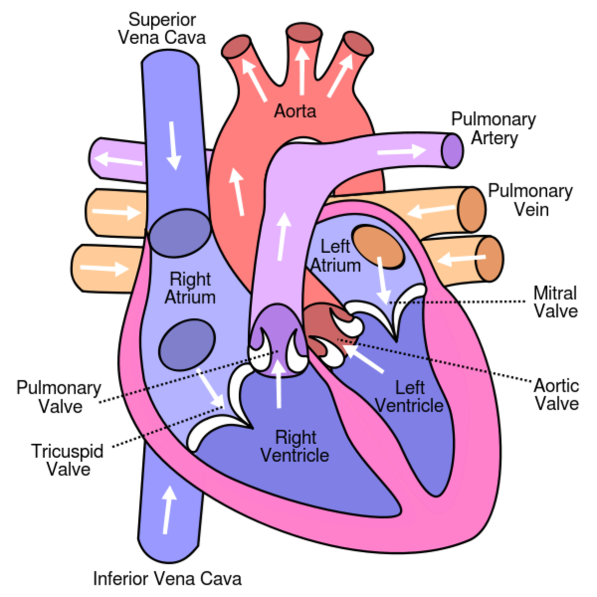
Heart Anatomy Draw - Web to draw the internal structure of the heart, start by sketching the 2 pulmonary veins to the lower left of the aorta and the bottom of the inferior vena cava slightly to the right of that. Web create a curved shape similar to an acorn or apple’s bottom half. The valves of the heart. This key circulatory system structure is comprised of four chambers. Xxxl very detailed human heart. Receives oxygenated blood from the lungs. January 29, 2024 | published on: The medical information on this site is provided as an information resource only, and is not to be used or relied on for any. 4.5k views 2 years ago scientific illustration | adobe. The heart has five surfaces: Muscle and tissue make up this powerhouse organ. It also has several margins: After all, we know that stress is bad for the heart! Web create a curved shape similar to an acorn or apple’s bottom half. Part of the teachme series. Carries deoxygenated blood to the lungs. Within the triangle, draw a horizontal and vertical centerline to split the triangle into four pieces. This tool provides access to several medical illustrations, allowing the user to interactively discover heart anatomy. After all, we know that stress is bad for the heart! Rotate the 3d model to see how the heart's valves control. Web browse 866 heart anatomy drawing photos and images available, or start a new search to explore more photos and images. This website is aimed to be an easily accessible web based app that features an on demand high fidelity rendering of the human heart to use while on rounds, in teaching conferences, or by bedside. Do you want a. Web medically reviewed by the healthline medical network — by the healthline editorial team — updated on january 20, 2018. Region of the heart that pumps oxygenated blood to the body. The valves of the heart. Receives oxygenated blood from the lungs. Do you want a fun way to learn the structure of the heart? Base (posterior), diaphragmatic (inferior), sternocostal (anterior), and left and right pulmonary surfaces. Do you want a fun way to learn the structure of the heart? The right margin is the small section of the right atrium that extends between the superior and inferior vena cava. Flaps that prevent backflow of blood. View heart anatomy drawing videos. Within the triangle, draw a horizontal and vertical centerline to split the triangle into four pieces. Then, fill in the base of the heart with the right and left ventricles and the right and left atriums. The valves of the heart. The medical information on this site is provided as an information resource only, and is not to be used. Within the triangle, draw a horizontal and vertical centerline to split the triangle into four pieces. [1] the main shape will be the basis for the left and right ventricles. Web medically reviewed by the healthline medical network — by the healthline editorial team — updated on january 20, 2018. Carries deoxygenated blood to the lungs. Your heart is a. Base (posterior), diaphragmatic (inferior), sternocostal (anterior), and left and right pulmonary surfaces. The heart is a mostly hollow, muscular organ composed of cardiac muscles. Angle the slightly tampered end of the shape to the left about 120 degrees. 4.5k views 2 years ago scientific illustration | adobe. Web to draw the internal structure of the heart, start by sketching the. Web in this lecture, dr mike shows the two best ways to draw and label the heart! It also has several margins: Carries deoxygenated blood to the lungs. The valves of the heart. Web browse 866 heart anatomy drawing photos and images available, or start a new search to explore more photos and images. Web carries deoxygenated blood from the body to the heart. Images are labelled, providing an invaluable medical and anatomical tool. The medical information on this site is provided as an information resource only, and is not to be used or relied on for any. This tool provides access to several medical illustrations, allowing the user to interactively discover heart anatomy.. After all, we know that stress is bad for the heart! Improve your drawing skills with printable practice sheets! Rotate the 3d model to see how the heart's valves control blood flow between heart chambers and blood flow out of the heart. Flaps that prevent backflow of blood. Draw the main shape of your human heart drawing. Carries deoxygenated blood to the lungs. [1] the main shape will be the basis for the left and right ventricles. Web carries deoxygenated blood from the body to the heart. Web function and anatomy of the heart made easy using labeled diagrams of cardiac structures and blood flow through the atria, ventricles, valves, aorta, pulmonary arteries veins, superior inferior vena cava, and chambers. New 3d rotate and zoom. Web in this lecture, dr mike shows the two best ways to draw and label the heart! The heart has five surfaces: January 29, 2024 | published on: Web create a curved shape similar to an acorn or apple’s bottom half. Web anatomy of the human heart and coronaries: The heart is a mostly hollow, muscular organ composed of cardiac muscles.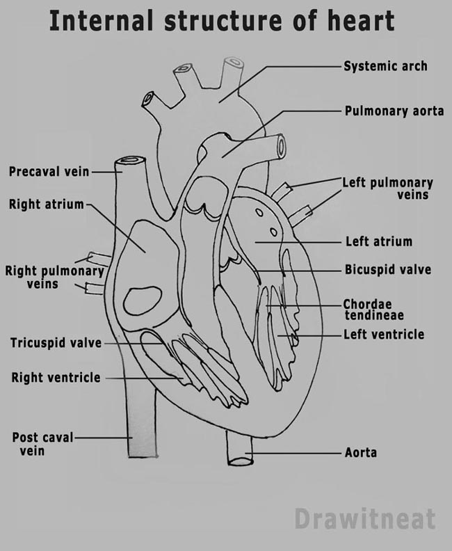
DRAW IT NEAT How to draw human heart labeled

How to Draw the Internal Structure of the Heart (with Pictures)

How to draw Structure of heart and blood circulation drawing step by

How To Draw Human Heart Diagram

How to Draw the Internal Structure of the Heart 13 Steps
Anatomical Drawing Heart at GetDrawings Free download

Heart Diagram Sketch at Explore collection of
How to draw Human Heart with colour Human Heart labelled diagram
Module 13 Heart and Great Vessels Anatomy 337 eReader
Learn About the Heart and Circulatory System for Kids HubPages
Receives Oxygenated Blood From The Lungs.
It’s Your Circulatory System ’S Main Organ.
Xxxl Very Detailed Human Heart.
Web How To Draw A Human Heart.
Related Post: