Golgi Drawing
Golgi Drawing - Web figure 2 shows golgi's drawing of the cell types of the cerebellar cortex. In the legend golgi stated that the plate, illustrating “some types of ganglion cells in the cerebral cortex,” is especially destined to show the origin and branching of the axon (“the only nervous prolongation of. The black stained cells in the molecular layer are basket cells. Golgi believed that basket cell axons continue on past the purkinje cell body to end in an axonal net within the granular layer. Protoplasmic prolongations branch dichotomously in. Web because of the great contrast between cell and background, every single part of the neuron was completely filled, allowing golgi to do drawings of the morphology of this nervous tissue. Web drawing by camillo golgi of a hippocampus stained with the silver nitrate method. Early illustrations of the nervous system by camillo golgi and santiago ramón y cajal. Web golgi's method is a silver staining technique that is used to visualize nervous tissue under light microscopy. The method was discovered by camillo golgi, an italian physician and scientist, who published the first picture made with the technique in 1873. The pale brown ovals are the cell bodies of the purkinje cell. In the legend golgi stated that the plate, illustrating “some types of ganglion cells in the cerebral cortex,” is especially destined to show the origin and branching of the axon (“the only nervous prolongation of. Web golgi’s first published drawing of neurons stained using the black reaction featured. Golgi used a camera lucida to project an image of the stained tissue onto paper so. Nerve cells in a dog's olfactory bulb (detail), from camillo golgi's sulla fina anatomia degli organi centrali del sistema nervoso (1885) —. The black stained cells in the molecular layer are basket cells. Web golgi's method is a silver staining technique that is used. Golgi believed that basket cell axons continue on past the purkinje cell body to end in an axonal net within the granular layer. In the legend golgi stated that the plate, illustrating “some types of ganglion cells in the cerebral cortex,” is especially destined to show the origin and branching of the axon (“the only nervous prolongation of. Early illustrations. Protoplasmic prolongations branch dichotomously in. The black stained cells in the molecular layer are basket cells. Based on his staining results, golgi supported the idea that the parts of the nervous system are all one very large, physically connected network. Web drawing by camillo golgi of a hippocampus stained with the silver nitrate method. Web because of the great contrast. Web because of the great contrast between cell and background, every single part of the neuron was completely filled, allowing golgi to do drawings of the morphology of this nervous tissue. Web golgi’s first published drawing of neurons stained using the black reaction featured the nervous structures of a dog’s olfactory bulb. Golgi used a camera lucida to project an. Based on his staining results, golgi supported the idea that the parts of the nervous system are all one very large, physically connected network. Web indeed, golgi's drawing was specifically made to show shapes, ramification laws, dispositions, localisation, and relationships of the large ganglionic cells of the granular layer. (a) the drawing is plate i from golgi (1885). Web golgi's. Based on his staining results, golgi supported the idea that the parts of the nervous system are all one very large, physically connected network. Nerve cells in a dog's olfactory bulb (detail), from camillo golgi's sulla fina anatomia degli organi centrali del sistema nervoso (1885) —. The black stained cells in the molecular layer are basket cells. Early illustrations of. Nerve cells in a dog's olfactory bulb (detail), from camillo golgi's sulla fina anatomia degli organi centrali del sistema nervoso (1885) —. Web after publishing a series of papers with descriptions and drawings of previously unseen structures such as the brain’s hippocampus and olfactory bulb, made visible by his silver staining technique, golgi was appointed professor of histology at. In. Web indeed, golgi's drawing was specifically made to show shapes, ramification laws, dispositions, localisation, and relationships of the large ganglionic cells of the granular layer. Protoplasmic prolongations branch dichotomously in. (a) the drawing is plate i from golgi (1885). Golgi believed that basket cell axons continue on past the purkinje cell body to end in an axonal net within the. Protoplasmic prolongations branch dichotomously in. Web golgi’s first published drawing of neurons stained using the black reaction featured the nervous structures of a dog’s olfactory bulb. Web after publishing a series of papers with descriptions and drawings of previously unseen structures such as the brain’s hippocampus and olfactory bulb, made visible by his silver staining technique, golgi was appointed professor. Web golgi’s first published drawing of neurons stained using the black reaction featured the nervous structures of a dog’s olfactory bulb. Based on his staining results, golgi supported the idea that the parts of the nervous system are all one very large, physically connected network. Web drawing by camillo golgi of a hippocampus stained with the silver nitrate method. The method was discovered by camillo golgi, an italian physician and scientist, who published the first picture made with the technique in 1873. The pale brown ovals are the cell bodies of the purkinje cell. Nerve cells in a dog's olfactory bulb (detail), from camillo golgi's sulla fina anatomia degli organi centrali del sistema nervoso (1885) —. Web after publishing a series of papers with descriptions and drawings of previously unseen structures such as the brain’s hippocampus and olfactory bulb, made visible by his silver staining technique, golgi was appointed professor of histology at. The black stained cells in the molecular layer are basket cells. Web indeed, golgi's drawing was specifically made to show shapes, ramification laws, dispositions, localisation, and relationships of the large ganglionic cells of the granular layer. Golgi believed that basket cell axons continue on past the purkinje cell body to end in an axonal net within the granular layer. Golgi used a camera lucida to project an image of the stained tissue onto paper so. (a) the drawing is plate i from golgi (1885). Web because of the great contrast between cell and background, every single part of the neuron was completely filled, allowing golgi to do drawings of the morphology of this nervous tissue. Early illustrations of the nervous system by camillo golgi and santiago ramón y cajal. In the legend golgi stated that the plate, illustrating “some types of ganglion cells in the cerebral cortex,” is especially destined to show the origin and branching of the axon (“the only nervous prolongation of.
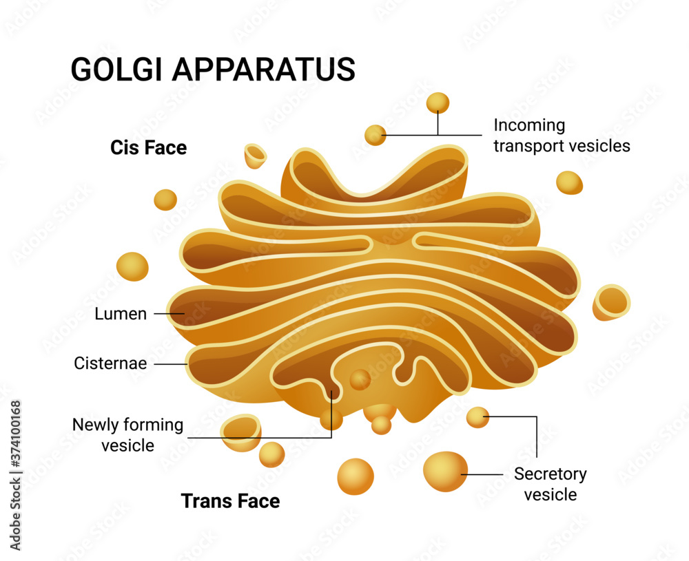
Structure and function of the golgi apparatus
Vector Illustration of a Golgi apparatus structure. Educational

How TO Draw golgi body easy/golgi apparatus drawing easy YouTube
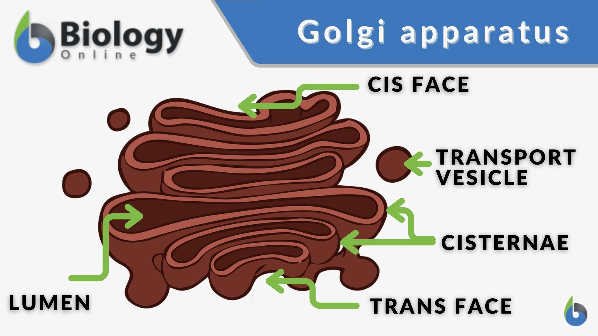
Golgi apparatus Definition and Examples Biology Online Dictionary

Structure and function of the golgi apparatus

How TO Draw golgi body apparatus/diagram of golgi body/golgi body

How TO Draw golgi body step by step/golgi body drawing YouTube
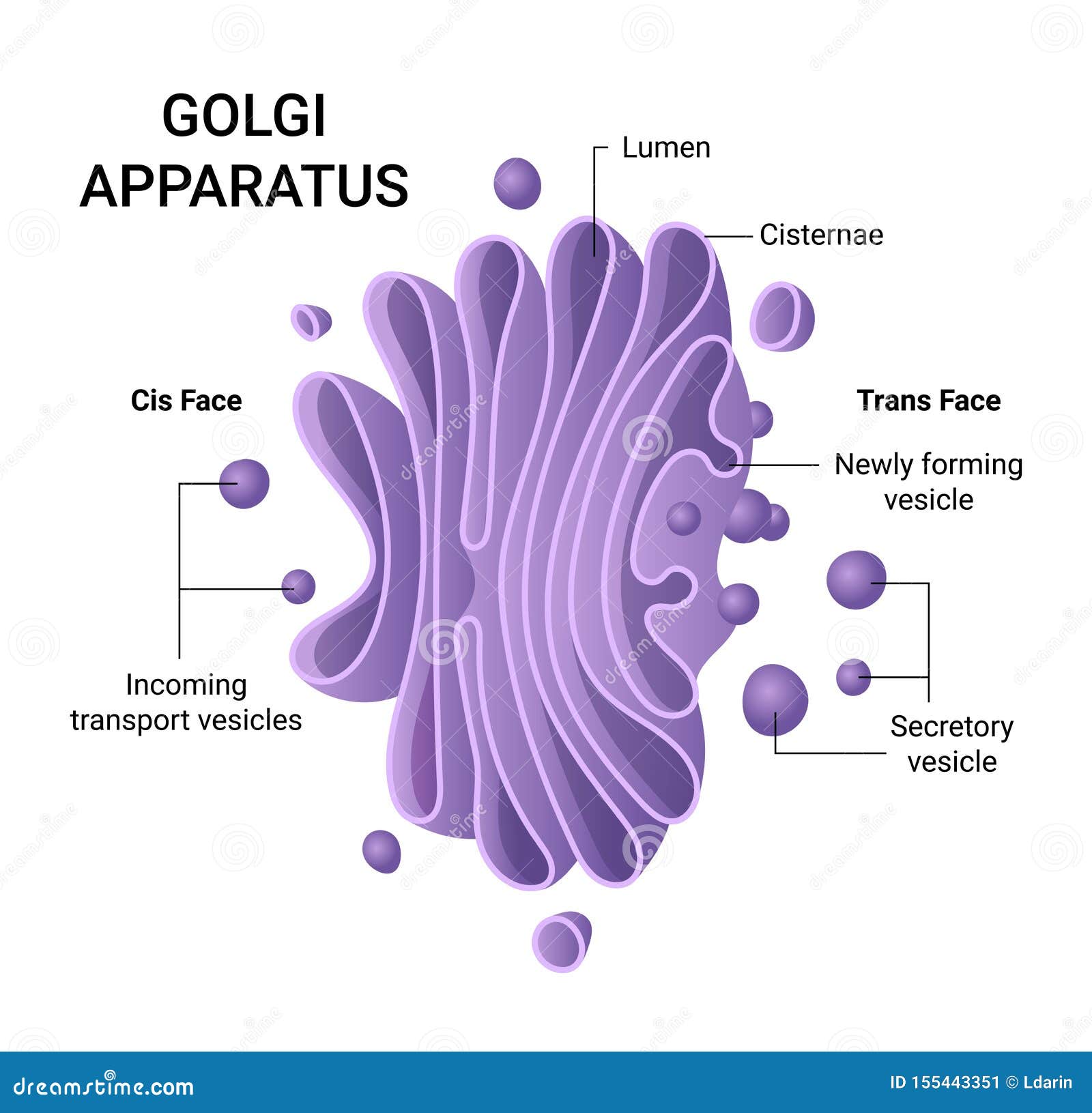
Illustration of the Golgi Apparatus Structure. Vector Infographics

Golgi Apparatus Structure Drawing Biology4kids com cell structure golgi

how to draw diagram of golgi body easily, YouTube
Web Figure 2 Shows Golgi's Drawing Of The Cell Types Of The Cerebellar Cortex.
Web Golgi's Method Is A Silver Staining Technique That Is Used To Visualize Nervous Tissue Under Light Microscopy.
Protoplasmic Prolongations Branch Dichotomously In.
In 1871, A German Anatomist Joseph Von Gerlach Postulated That The Brain Is A Complex Protoplasmic Network, In The Form Of A Continuous Network Called The Reticulum.
Related Post: