Drawing Uterus
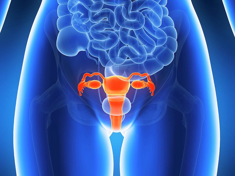
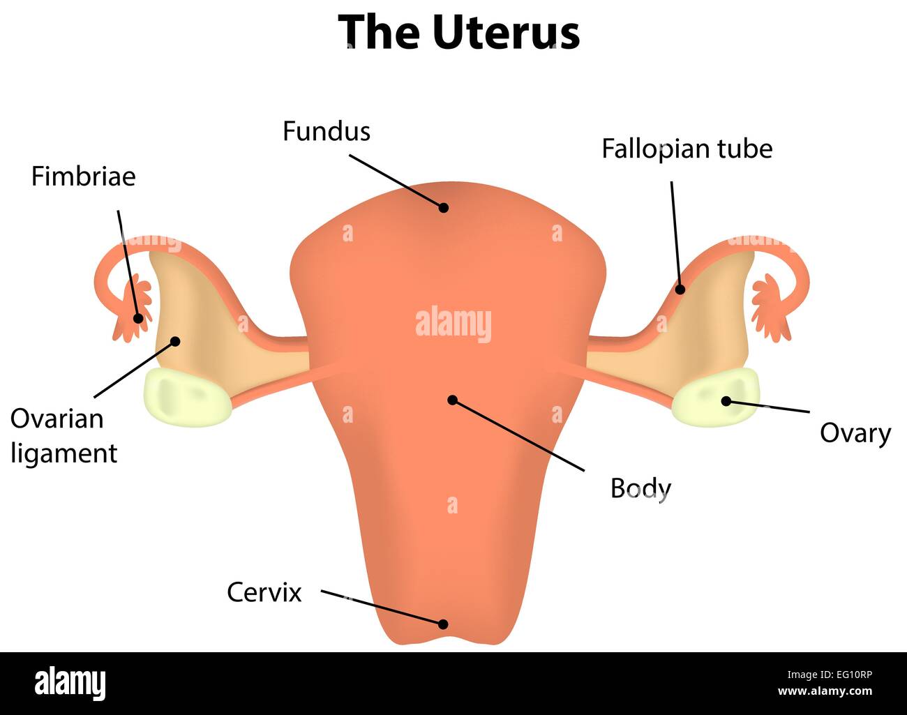
Drawing Uterus - The uterus is where a fetus develops during pregnancy. Web by alice dreger updated may 3, 2024, 3:00 a.m. The lining of the uterus. It is located posterior to the urinary bladder and is connected via the cervix to the vagina on its inferior border and the fallopian tubes along its superior end. The vulva and its structures form the external genitalia. Web anatomy of the female reproductive system; Web the uterus is approximately the shape and size of a pear and sits in an inverted position within the pelvic cavity of the torso. A beautiful drawing of the female. White blood cells are extracted from a blood. Web the uterus opens into the vagina via the cervix. The cervix and the corpus. How you will use this image and then you will be able to add this image to. Your uterus is divided into two parts: The female reproductive system includes the ovaries, fallopian tubes, uterus, and vagina. The uterus has three layers of muscle and is one of the strongest muscles in the body. This part is structurally and functionally different to the rest of the uterus. 42k views 3 years ago. How you will use this image and then you will be able to add this image to. It is located along the body’s midline posterior to the urinary bladder and anterior to the rectum. The function of the uterine cervix during pregnancy. Your uterus is divided into two parts: It is usually present in. The female reproductive system includes the ovaries, fallopian tubes, uterus, and vagina. How you will use this image and then you will be able to add this image to. Web histology of the uterus. These tubes help the uterus to communicate with the peritoneal cavity. The uterus is an organ in a person's pelvis. It's also called the womb. A beautiful drawing of the female. The lining of the uterus. The female reproductive system includes the ovaries, fallopian tubes, uterus, and vagina. These tubes help the uterus to communicate with the peritoneal cavity. The uterus is where a fetus develops during pregnancy. Web histology of the uterus. (realistic specimen) • demo uterus. Two female reproductive organs located in the pelvis. Web histology of the uterus. The uterus has three layers of muscle and is one of the strongest muscles in the body. The vulva and its structures form the external genitalia. Web the uterus has three parts; The lining of the uterus. The female reproductive system includes the ovaries, fallopian tubes, uterus, and vagina. This short article describes the normal anatomy of the uterus and will focus on definitions, structure, location, supporting ligaments, blood supply and innervation. It's also called the womb. The uterus is where a fetus develops during pregnancy. The endometrium (uterine mucous membrane) is lined with simple columnar epithelium (lamina epithelialis) and contains numerous tubular glands. Web the uterus opens into the vagina via the cervix. It's also called the womb. It is located posterior to the urinary bladder and is connected via the cervix to the vagina on its inferior border and the fallopian tubes along its. Web uterus, ovaries, and uterine tubes variant image id: It is located posterior to the urinary bladder and is connected via the cervix to the vagina on its inferior border and the fallopian tubes along its superior end. Your uterus is divided into two parts: White blood cells are extracted from a blood. Web the female reproductive system includes external. The uterine tubes, the uterus, and the vagina. Drawing shows the uterus, myometrium (muscular outer layer of the uterus), endometrium (inner lining of the uterus), ovaries, fallopian tubes, cervix, and vagina. Carry eggs from the ovaries to the uterus. Web anatomy of the female reproductive system; White blood cells are extracted from a blood. It’s where a fertilized egg implants during pregnancy and where your baby develops until birth. A beautiful drawing of the female. Web anatomy of the female reproductive system; Drawing shows the uterus, myometrium (muscular outer layer of the uterus), endometrium (inner lining of the uterus), ovaries, fallopian tubes, cervix, and vagina. Web the uterus is approximately the shape and size of a pear and sits in an inverted position within the pelvic cavity of the torso. It's where a fetus (unborn baby) develops and grows during pregnancy. Web uterus, ovaries, and uterine tubes variant image id: Web the uterus has three parts; It is located posterior to the urinary bladder and is connected via the cervix to the vagina on its inferior border and the fallopian tubes along its superior end. Web histology of the uterus. This short article describes the normal anatomy of the uterus and will focus on definitions, structure, location, supporting ligaments, blood supply and innervation. Semenya was born intersex and has. Browse 668 uterus drawing photos and images available, or search for uterus illustration to find more great photos and pictures. The cervix and the corpus. The female reproductive organs include several key structures, such as the ovaries, uterus, vagina, and. The ovaries produce eggs and hormones, and the fallopian tubes.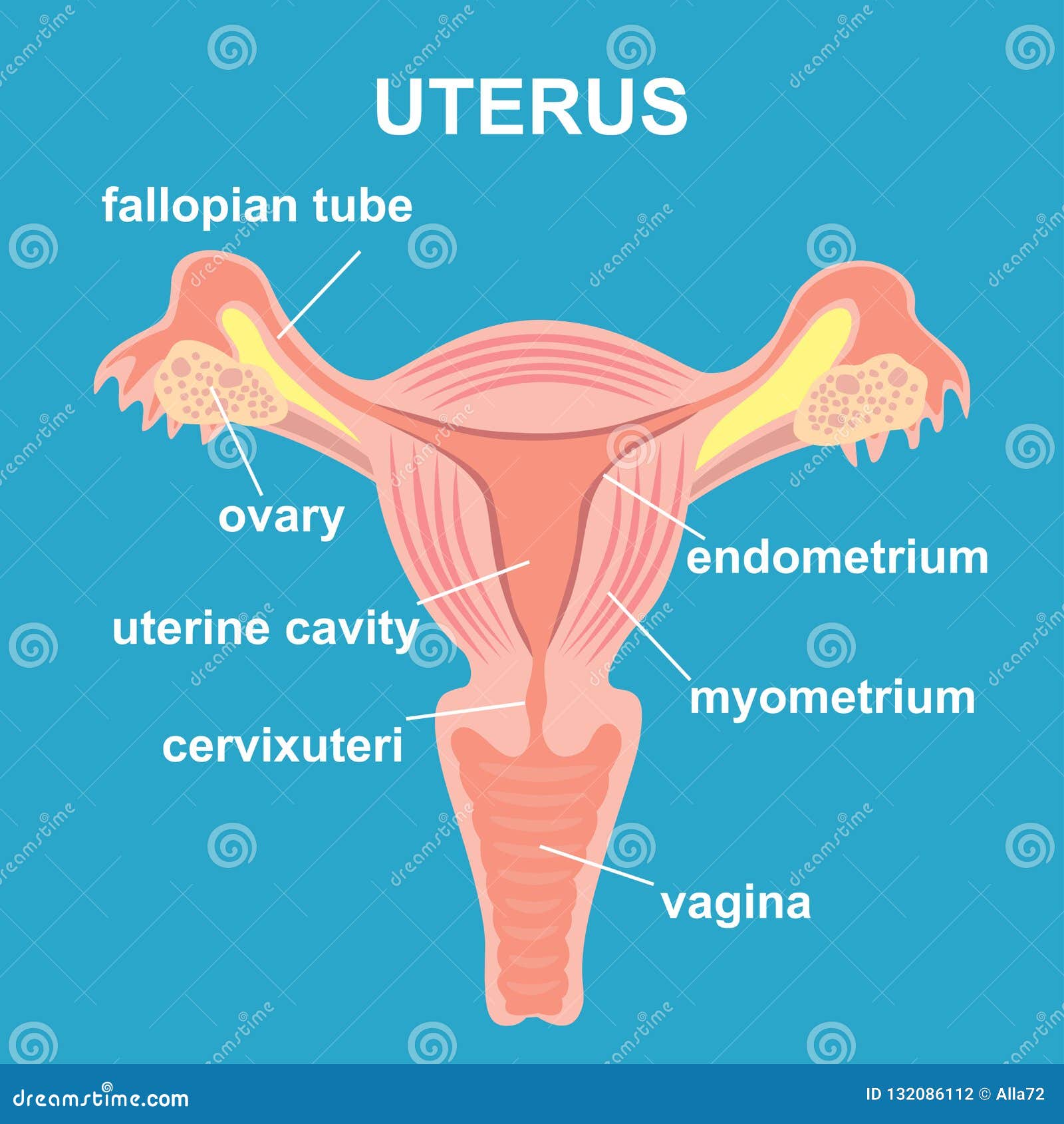
Uterus and Ovaries, Organs of Female Reproductive System Stock Vector
![]()
Uterus. Realistic handdrawn icon of human internal organs. Engraving
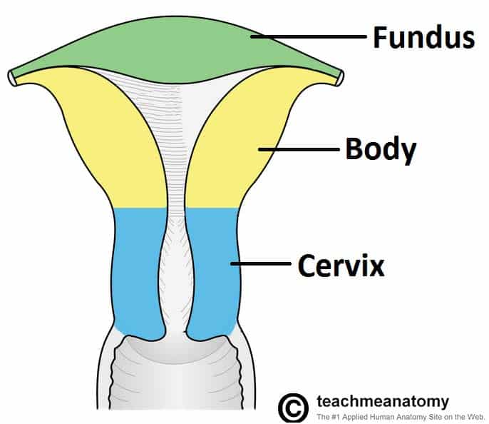
The Uterus Structure Location Vasculature TeachMeAnatomy
Uterus Anatomy Uterus Women's Health

Uterus, Ovaries, Fallopian Tubes, Illustration Stock Image C043
Anatomy Of Human Uterus Photograph by Sebastian Kaulitzki Fine Art
Uterus Labeled Diagram Stock Vector Image & Art Alamy

How to draw a Uterus parts of the inner body Easy step by step

Contour Anatomical Sketch of the Uterus. Healthy Female Body. Woman

dibujo de arte de línea continua del útero reproductivo femenino
How You Will Use This Image And Then You Will Be Able To Add This Image To.
Web Anatomy Atlas Of The Female Pelvis:
Web The Uterus, Also Known As The Womb, Is A Hollow, Muscular Organ Located In The Pelvis Between The Bladder And Rectum.
This System Of Ducts Connects To The Ovaries, The Primary Reproductive Organs.
Related Post:
