Drawing Of The Eye Anatomy
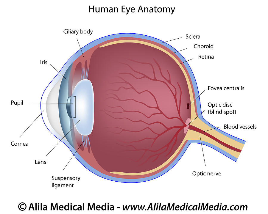
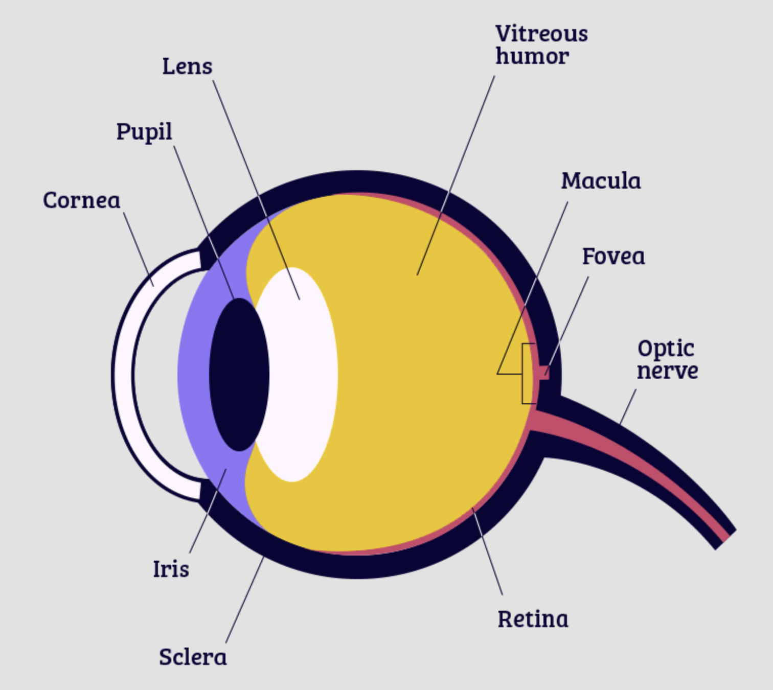
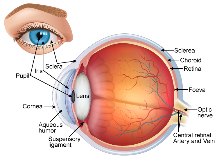
Drawing Of The Eye Anatomy - Web human eye anatomy (seen from above) for more details about specific structures of the eye and how they function, visit these pages: Santiago ramón y cajal, 1904. External landmarks and extraocular muscles. Web the eye's structure includes the sclera, cornea, conjunctiva, aqueous humour, lens, ciliary body, iris, pupil, vitreous humour, retina, optic nerve, choroid, fovea, and macula. Study the diagram below or click here for an interactive study guide and game! Curved to bend light into your eye, its tough and clear like a windshield to protect your eye from dust. Quiz on the 5 layers of the cornea. 442k views 3 years ago anatomy for artists. The bottom panel shows inside of the eye including the cornea, lens, ciliary body, retina, choroid, optic nerve, and vitreous humor. The eye is the organ that allows sight. Only the most important anatomical details are listed, alongside web links to videos and diagrams (each underlined text is a link) external eye and adnexa. Pick the right drawing tools. Web learning artistic anatomy for drawing the eye? Web in this tutorial i cover how to draw the structure of the eye and it’s anatomy. Dec 26, 2023 3:25 pm. We explore the proportional relationships between the eye lids, the eye socket, the eyeball, and more. Eyes are often the first point of contact when meeting a person. Below, find an explanation of each anatomical part from the above video with physiologic and pathologic correlates. Jana vasković, md • reviewer: Instead, it is made up of two separate segments fused. Web in this tutorial i cover how to draw the structure of the eye and it’s anatomy. Curved to bend light into your eye, its tough and clear like a windshield to protect your eye from dust. Quiz on the 5 layers of the cornea. Web reviewed by ninel z gregori, md. Web eye anatomy (16 parts of the eye. Anatomy of the human eye. Pick the right drawing tools. Anterior chamber angle and ciliary body. Eyeball [25:37] structure of the eyeball seen in a transverse section. For more video tutorials visit www.proko.com and subscribe to the newsletter. Anterior chamber angle and ciliary body. The eyeball, eye socket, brow ridge, eyelids, tear duct, sclera, iris, pupil, cornea, glabella, and epicanthic fold. The bottom panel shows inside of the eye including the cornea, lens, ciliary body, retina, choroid, optic nerve, and vitreous humor. Web reviewed by ninel z gregori, md. These interactive figures are provided for use in medical. Bhavin shah, neurodevelopmental and behavioral optometrist specializing in myopia management, central vision opticians. Below, find an explanation of each anatomical part from the above video with physiologic and pathologic correlates. The white of the eye. Web the eye's structure includes the sclera, cornea, conjunctiva, aqueous humour, lens, ciliary body, iris, pupil, vitreous humour, retina, optic nerve, choroid, fovea, and macula.. Jana vasković, md • reviewer: Bhavin shah, neurodevelopmental and behavioral optometrist specializing in myopia management, central vision opticians. Web structure and functions. Here is a tour of the eye starting from the outside, going in through the front and working to the back. Eyes are often the first point of contact when meeting a person. For more video tutorials visit www.proko.com and subscribe to the newsletter. Anterior chamber angle and ciliary body. Jana vasković, md • reviewer: We explore the proportional relationships between the eye lids, the eye socket, the eyeball, and more. Get to know the eye structure. The bottom panel shows inside of the eye including the cornea, lens, ciliary body, retina, choroid, optic nerve, and vitreous humor. Web in this tutorial i cover how to draw the structure of the eye and it’s anatomy. Web learning artistic anatomy for drawing the eye? “cajal’s drawings of the retina are as beautiful as they are anatomically accurate. Eyeball. Below, find an explanation of each anatomical part from the above video with physiologic and pathologic correlates. For more video tutorials visit www.proko.com and subscribe to the newsletter. Web human eye anatomy (seen from above) for more details about specific structures of the eye and how they function, visit these pages: Web eye anatomy (16 parts of the eye &. Here is a tour of the eye starting from the outside, going in through the front and working to the back. 442k views 3 years ago anatomy for artists. Study the diagram below or click here for an interactive study guide and game! External landmarks and extraocular muscles. Contrary to popular belief, the eyes are not perfectly spherical; The eyeball, eye socket, brow ridge, eyelids, tear duct, sclera, iris, pupil, cornea, glabella, and epicanthic fold. Eyeball (bulbus oculi) the eye is a highly specialized sensory organ located within the bony orbit. It's made up of many parts—each with specific names and functions. Get to know the eye structure. Full and extensive eye anatomy in drawing for artists. Santiago ramón y cajal, 1904. Web the eye's structure includes the sclera, cornea, conjunctiva, aqueous humour, lens, ciliary body, iris, pupil, vitreous humour, retina, optic nerve, choroid, fovea, and macula. A hole in the middle of the iris that changes size to let in more or less light. Blood supply of the eye. Pick the right drawing tools. Quiz on the 5 layers of the cornea.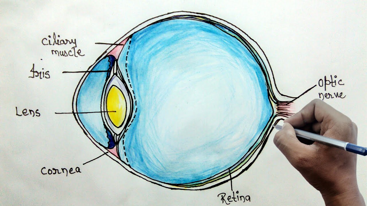
How to draw human eye diagram for beginners YouTube

File1413 Structure of the Eye.jpg Wikimedia Commons
Eye Anatomy Labeled Drawing
About the Eye National Eye Institute

Anatomy of the Human Eye
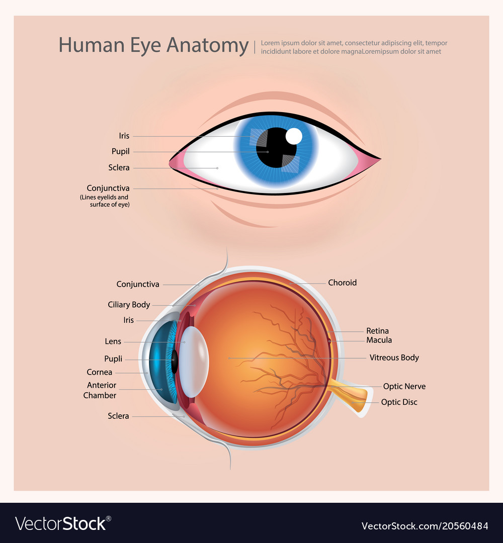
Human eye anatomy Royalty Free Vector Image VectorStock
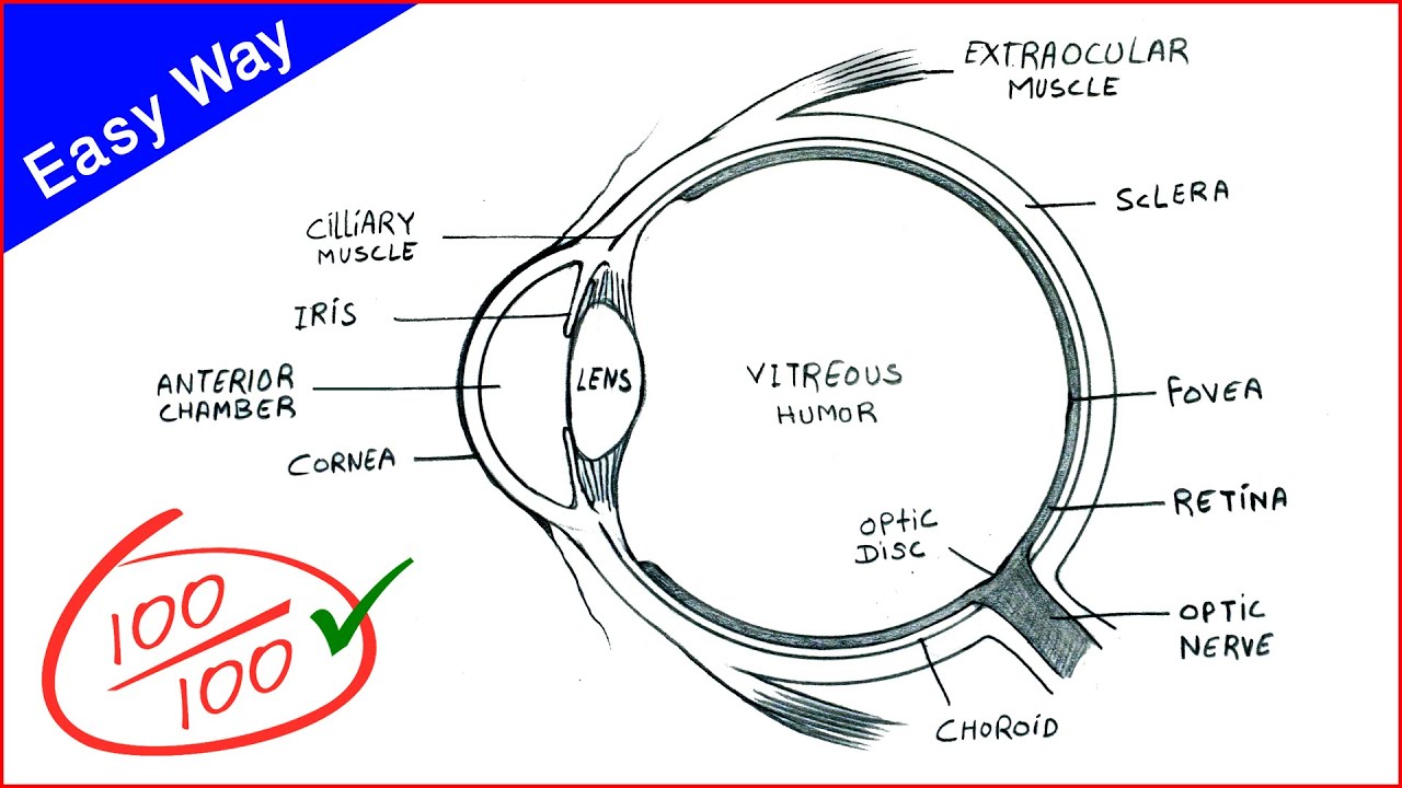
Eye Diagram drawing CBSE easy way draw Human eye anatomy Step

Diagram human eye anatomy with label Royalty Free Vector
draw a neat and labelled diagram of structure of the human eye slwbyx77

Labeled Simple Labeled Human Eye Diagram
To Understand The Diseases And Conditions That Can Affect The Eye, It Helps To Understand Basic Eye Anatomy.
Web Structure And Functions.
318 Views 1 Year Ago Scientific Diagram Drawing Tutorials.
Discussion Led By Art Prof Clara Lieu.
Related Post: