Amoeba Cell Drawing
Amoeba Cell Drawing - Amoebae are a type of eukaryotic organism made up of. Typically belonging to the kingdom protozoa, it moves in an “amoeboid” fashion. Amoebae are microscopic unicellular organisms that belong to the family amoebidae. Some parasitic amoebae living inside animal bodies, including. This page is a draft and is under active development. Amoebas can form temporary extensions of their cytoplasm known as pseudopodia or false feet which can be used for locomotion or capturing food. They have a movable shape and can extend pseudopods to reach for and engulf prey. Color the cellular structures of the ameba. Web amoeba cell diagram. They can be identified by their shape changing ability from their cytoplasmic extensions called pseudopodia. This will also help you to draw the structure and diagram of amoeba. Color the cellular structures of the ameba. They prey on smaller organisms, such as bacteria. The ameba (also spelled amoeba) is a protozoan that belongs to the kingdom protista. Using the definitions listed below, label the amoeba. Web amoeba proteus is a microscopic living organism which consists of a single cell. They prey on smaller organisms, such as bacteria. Like all cells, it has cytoplasm, nucleus, cell membrane and a variety of inclusions in the cytoplasm. The ameba (also spelled amoeba) is a protozoan that belongs to the kingdom protista. A sample of water collected from a. Amoebae are microscopic unicellular organisms that belong to the family amoebidae. To view amoebas under the microscope, students will need the following: As such, microbiologists often use the term “amoeboid”, to refer to a specific type of movement and amoebae interchangeably. Web like an ordinary cell the body of amoeba has 3 main parts: 89k views 6 years ago. Fresh water and free living organism commonly available in stagnant water. Color the cellular structures of the ameba. An amoeba is a highly motile eukaryotic, unicellular organism. Web amoeba cell diagram. Amoebas can be found freely living and thriving in shallow pond waters with organic material. An amoeba is a highly motile eukaryotic, unicellular organism. Amoebas can be found freely living and thriving in shallow pond waters with organic material. Fresh water and free living organism commonly available in stagnant water. Plasma lemma or plasma membrane, cytoplasm and nucleus. It allows some substances to. They can be identified by their shape changing ability from their cytoplasmic extensions called pseudopodia. They have a movable shape and can extend pseudopods to reach for and engulf prey. A sample of water collected from a pond with organic material. Plant and animal cells venn diagram. It is capable of movement. Like all cells, it has cytoplasm, nucleus, cell membrane and a variety of inclusions in the cytoplasm. Using the definitions listed below, label the amoeba. Plant and animal cells venn diagram. Less commonly spelled ameba or amœba; Web amoebas are used extensively in cell research for determining the relative functions and interactions of the nucleus and the cytoplasm. Web by rebecca mayglothling. Some parasitic amoebae living inside animal bodies, including. They prey on smaller organisms, such as bacteria. Worksheet questions can be answered by reading the passage carefully. Fresh water and free living organism commonly available in stagnant water. The ameba (also spelled amoeba) is a protozoan that belongs to the kingdom protista. Even though a bacterium is just one cell, it can carry out all seven life processes. Color the cellular structures of the ameba. Art activities and projects for students. Amoebas (less commonly, amebas) or amoebae (amebae) / ə ˈ m iː b i /), often called. Typically belonging to the kingdom protozoa, it moves in an “amoeboid” fashion. Plasma lemma is a very thin, delicate and elastic cell membrane of amoeba. They have a movable shape and can extend pseudopods to reach for and engulf prey. Amoebae are microscopic unicellular organisms that belong to the family amoebidae. Web amoebas are used extensively in cell research for. This page is a draft and is under active development. Color the cellular structures of the ameba. Amoebae are microscopic unicellular organisms that belong to the family amoebidae. Some parasitic amoebae living inside animal bodies, including. Download a free printable outline of this video and draw along with us:. Art activities and projects for students. Color the cellular structures of the ameba. Humans, on the other hand, are multicellular because we have approximately 37 trillion cells! Each amoeba contains a small mass of jellylike cytoplasm, which is differentiated into a thin outer plasma membrane, a layer of stiff, clear ectoplasm just within the plasma membrane, and a central granular. Amoebae are a type of eukaryotic organism made up of. Typically belonging to the kingdom protozoa, it moves in an “amoeboid” fashion. The living things we could see are multicellular as we cannot see cells with our eyes. Plant and animal cells venn diagram. They have a movable shape and can extend pseudopods to reach for and engulf prey. When seen under a microscope, the cell looks like a tiny blob of colorless jelly with a dark speck inside it. Web by rebecca mayglothling.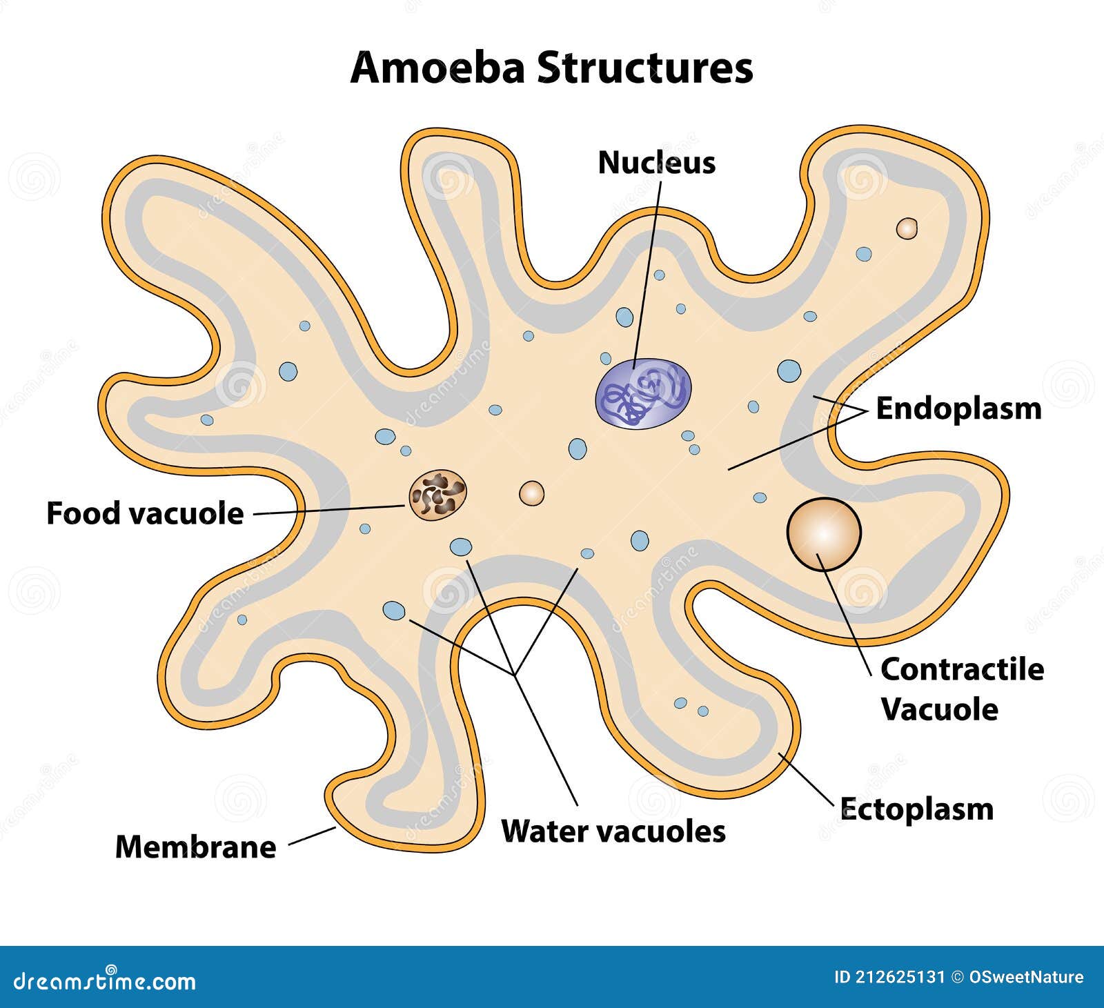
Amoeba Cell Structures Biology Diagram Stock Vector Illustration of
Structure of Amoeba in illustration. Contractile vacuole, and

Amoeba labeled vector illustration in 2022 Vector illustration

How TO Draw amoeba/easy way drawing amoeba/amoeba drawing YouTube
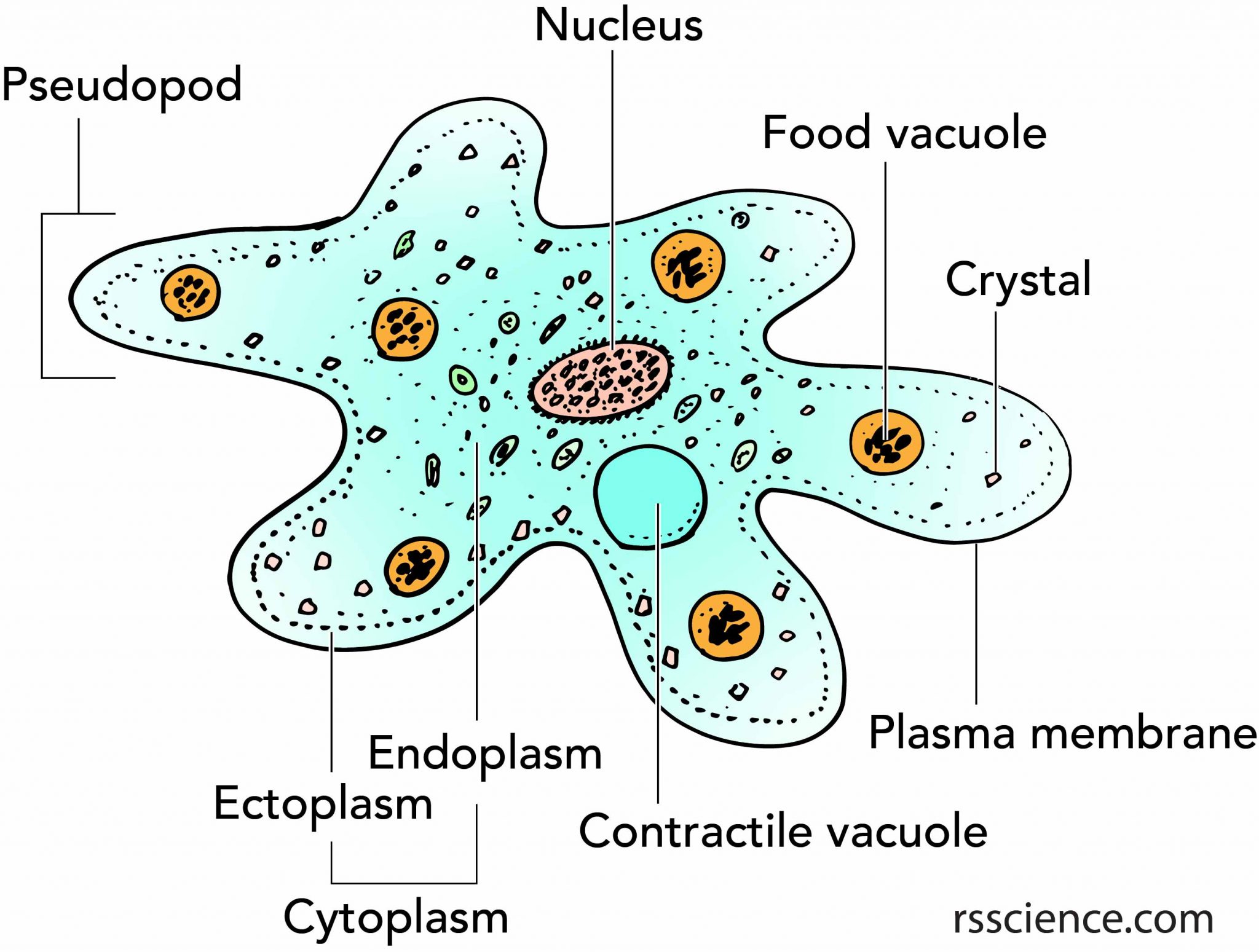
Facts about Amoeba, structure, behavior and reproduction Rs' Science
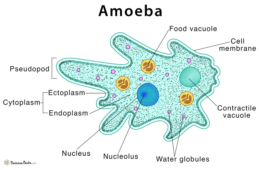
Amoeba Definition, Structure, & Characteristics with Diagram

How TO Draw amoeba step by step/amoeba drawing easy YouTube
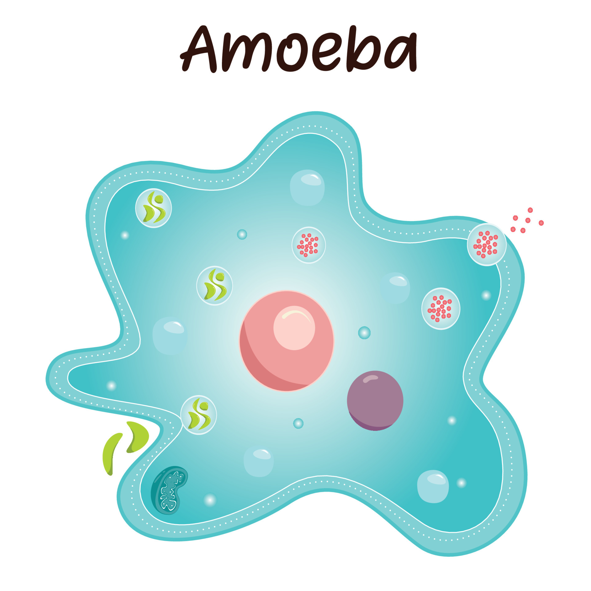
Diagram of an Amoeba 6787644 Vector Art at Vecteezy

Amoeba Labeled anatony of amoeba proteus, Detailed vector
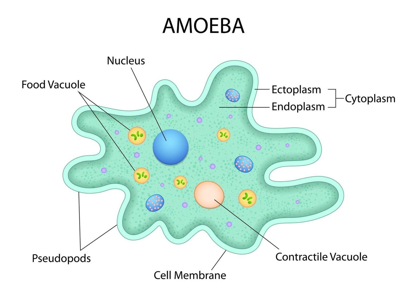
illustration of Healthcare and Medical education drawing chart of
©Sheri Amsel Www.exploringnature.org Food Vacuole Nucleus Contractile Vacuole.
Even Though A Bacterium Is Just One Cell, It Can Carry Out All Seven Life Processes.
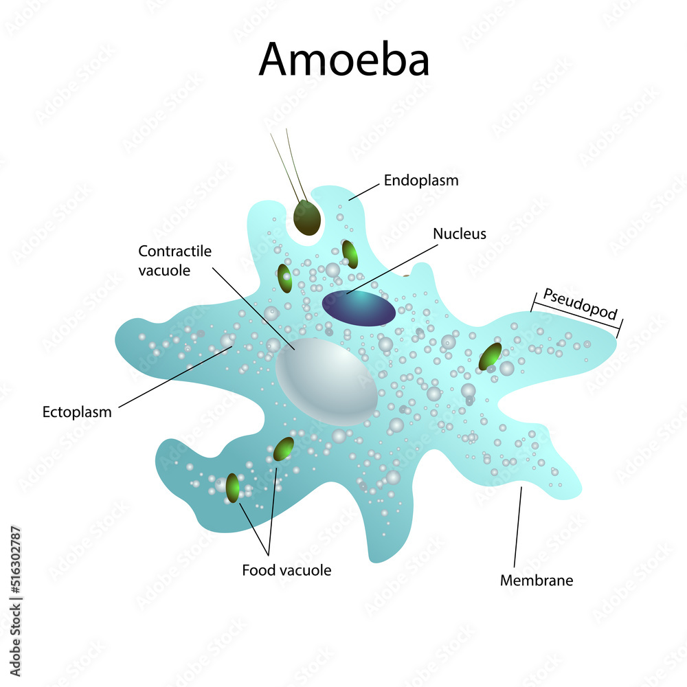
Web Like An Ordinary Cell The Body Of Amoeba Has 3 Main Parts:
It Can Be Dangerous Once It Infects A Human Host, Although By Observing Proper Safety Guidelines At Your Lab, It’ll Be Perfectly Fine To Observe And Experiment On These Microbes.
Related Post: